Abdominal ultrasound is an imaging examination primarily used to investigate a specific medical question. Ultrasound can be used to assess, among other things, the liver, gallbladder, bile ducts, kidneys, spleen and parts of the pancreas.
Abdominal ultrasound – for abdominal pain, swelling or abnormal test results
Abdominal ultrasound may, for example, be used when there is a suspicion of gallstones, changes in the liver or bile ducts, fluid in the abdomen or certain changes in the kidneys. It can also be used to follow up previously known findings in the abdomen.
Abdominal ultrasound is not a general screening examination and should not be used to check whether all organs in the abdomen are healthy. What can be assessed depends, among other things, on the underlying medical question, where in the abdomen the symptoms are located and need to be examined, and the anatomical conditions.
Abdominal ultrasound – for targeted medical questions
Abdominal ultrasound may be appropriate when symptoms or test results raise suspicion of a specific change in the abdomen. This may, for example, involve pain beneath the right rib cage with suspected gallstones, elevated liver test results, fluid in the abdomen or questions relating to the kidneys.
Common symptoms and medical questions
Abdominal ultrasound may, among other things, be used for:
- pain in the upper abdomen, for example when gallstones are suspected
- elevated liver test results or suspected changes in the liver or bile ducts
- suspected fluid accumulation in the abdomen, known as ascites
- suspected cysts or other localised changes in the liver or kidneys
- questions relating to the kidneys and urinary tract, for example suspected urinary obstruction
- follow-up of previous findings in, for example, the liver, kidneys or spleen
Symptoms such as abdominal pain, nausea and bloating can have many different causes. Ultrasound is therefore not always the appropriate examination based on a symptom alone. The choice of imaging method should be based on what specifically needs to be investigated.
What can be seen on an abdominal ultrasound?
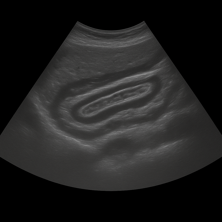
Ultrasound cannot rule out all types of disease or changes in the abdomen. Gas in the bowel, body build and the position of the organs can affect image quality. For some medical questions, CT or MRI may therefore be more appropriate. Abdominal ultrasound can, among other things, be used to assess:
- gallstones and certain changes in the gallbladder and bile ducts
- fatty changes, cysts and certain other changes in the liver
- fluid accumulation in the abdominal cavity
- cysts and certain structural changes in the kidneys
- dilatation of the renal pelvis or other signs of urinary obstruction
- enlargement or other structural changes in the spleen
- certain changes in the pancreas, when the organ can be visualised sufficiently well
How an abdominal ultrasound is performed
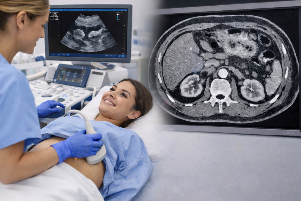
The examination is performed while you are lying on your back. Gel is applied to the skin and the doctor moves the ultrasound probe over the abdominal area to assess the internal organs. The examination takes approximately 20–30 minutes and is completely painless. For the best image quality, you should fast for 4–6 hours beforehand, as gas and food in the bowel can affect the images. If necessary, the urinary bladder may also be examined while full in order to assess urinary drainage from the kidneys.
Book an abdominal ultrasound – receive a report from a specialist doctor
The images are reviewed by a specialist doctor in radiology, who prepares a written medical report. The results are delivered digitally within a few working days and can be shared with your treating doctor for further follow-up or treatment. If necessary, findings may be followed up with MRI or CT for more detailed assessment.