Quick version
An X-ray or MRI scan describes what is seen in the image and is rarely a complete diagnosis in itself. Bone marrow edema indicates strain or irritation in the skeleton, while degenerative changes are completely normal wear and tear that does not always have to be painful. Fluid in the pelvis is often a natural part of the menstrual cycle, but symptoms such as bleeding or severe pain may require further assessment. The doctor therefore always weighs the images against how you actually feel, and emergency care should be sought in the event of sudden severe pain, fever or difficulty with exercise.
Bone marrow edema, degenerative changes and fluid in the pelvis are words that often appear in the results of an MRI, X-ray or ultrasound. For many, they come as a surprise: the examination may have been done for hip pain, knee problems or a dull ache in the lower abdomen, but the results contain terms that sound much more dramatic than they sometimes are. This is precisely why such findings need to be translated into standard Swedish.
What do findings such as bone marrow edema, degenerative changes and fluid in the pelvis mean?
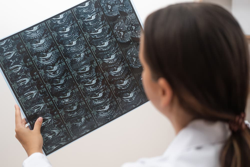
Bone marrow edema means that an increased amount of fluid is seen in the bone marrow cavity on an MRI. It is therefore an imaging finding, not a complete diagnosis in itself. It can be more simply thought of as the skeleton showing signs of strain, irritation or inflammation in the area.
This finding can occur in several different situations. Common causes are overload, stress fracture, changes similar to osteoarthritis, damage after trauma or certain inflammatory joint diseases. Sometimes bone marrow edema is also seen in more unusual conditions, such as transient bone marrow edema syndrome or osteonecrosis, which is why the finding must always be interpreted together with symptoms, age and where in the body the change is located.
For example, if you have pain in your hip when you walk and an MRI shows bone marrow edema in the femoral head, it may mean that the joint is severely irritated by stress or that the doctor needs to rule out another underlying cause. If the same finding is located near a knee with known arthrosis, it is often better to consider a stress-related change. The finding therefore says more that something is going on in the bone than exactly what the cause is.
Bone marrow edema often causes pain with stress and sometimes pain at rest. In some cases, the change heals with relief and time, but when the pain is clear or the finding is extensive, follow-up is needed to avoid missing, for example, a fracture, joint disease or reduced blood supply to the bone.
Degenerative changes – normal age-related wear and tear, but not always the whole explanation
Degenerative changes are a collective term for age- and load-related changes in joints, cartilage, discs, ligaments or bones. In practice, the term is often used when the radiologist sees signs that match osteoarthritis or other tissue changes that have developed gradually over time. This does not automatically mean that something is serious or that surgery is needed.
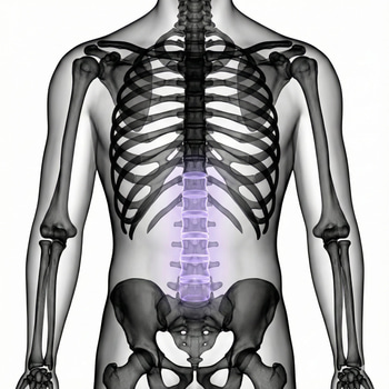
With osteoarthritis, the cartilage can become thinner, the bone changes and small bone spurs form. Such findings are common in, for example, the knee, hip, fingers and spine, especially with increasing age. Many people have degenerative changes on imaging without having any particular pain, while others have clear symptoms despite relatively mild findings.
That is why doctors do not assess an image in isolation. A 55-year-old with stiffness in the knee when standing up, pain when climbing stairs and degenerative changes on X-ray has a completely different situation than a person who has had an MRI after an acute trauma and where the same word happens to be listed as an incidental finding. The image should match what you actually feel in your body.
In cases of suspected osteoarthritis, symptoms and a medical examination are often enough. It is recommended that imaging diagnostics not be routinely used to diagnose osteoarthritis if the symptoms are typical, since images sometimes show more than what is clinically relevant.
Fluid in the pelvis – sometimes normal, sometimes a sign that must be put into the right context
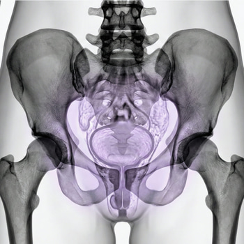
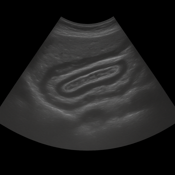
Fluid in the pelvis, or free fluid in the small pelvis, means that fluid has been seen in the abdominal cavity near the uterus, ovaries or intestine. In people with a menstrual cycle, a small amount of fluid can be completely physiological, especially in connection with ovulation or after a harmless functional cyst has ruptured.
However, the same finding can also be seen in gynecological problems. Examples are bleeding or ruptured ovarian cysts, endometriosis, pelvic inflammatory disease or early pregnancy complications such as ectopic pregnancy. Therefore, the amount of fluid, whether it looks clear or blood-tinged, and what symptoms you have at the same time are absolutely crucial for how the finding should be evaluated.
A common scenario is that someone is seeking treatment for unilateral pain in the lower abdomen and an ultrasound shows a small cyst plus some free fluid. If the person is otherwise well, this may indicate a cyst that has leaked or ruptured and which often resolves on its own. But if the same findings are seen in someone with missed periods, bleeding and increasing pain, ectopic pregnancy must be ruled out urgently.
In people without gynecological organs, or after menopause, fluid in the pelvis often has a different meaning and needs to be evaluated based on the entire medical picture. Then the doctor can think more about, for example, inflammation, surgical conditions or other fluid accumulation in the abdomen. This is another example of how the same words in a statement can mean different things in different bodies.
When are such findings harmless – and when should they be taken more seriously?
Many imaging findings are benign or at least not acute. Small degenerative changes in the back in a middle-aged person, a small amount of free pelvic fluid around ovulation or limited bone marrow edema after obvious overload are examples where the explanation is often relatively mundane. However, this does not mean that you should ignore problems that get worse or do not go away.
Warning signs are especially present when the findings are combined with clear symptoms. Seek prompt care if:
- sudden or very severe pain in the hip, knee or pelvis
- inability to bear weight on the leg
- fever, general ill health or suspected infection
- numbness, weakness or bladder or bowel problems with back pain
- missed period, suspected pregnancy, vaginal bleeding and simultaneous abdominal or pelvic pain
- feeling faint, cold sweats or rapidly increasing abdominal pain
In the case of long-standing but less acute problems, follow-up is often about linking the finding to the right next step. This may include physiotherapy for osteoarthritis and stress-related pain, unloading and a new check-up if a stress reaction in the skeleton is suspected, or a gynecological assessment if fluid in the pelvis is seen together with menstrual disorders or recurring pain.
What can you do yourself while the investigation is ongoing?
The first step is not to interpret the radiology report as a final diagnosis. The words in the report describe what is seen in the image, but the doctor needs to consider how much pain you have, where the pain is located, how long the problem has lasted and whether there are other symptoms such as fever, stiffness, bleeding or night pain.
If you have joint or bone pain, it is often helpful to note when the pain occurs: when straining, at rest or at night. This information makes it easier to distinguish between, for example, osteoarthritis, overload, inflammation and more acute conditions. In the case of pelvic pain, the connection to the menstrual cycle, ovulation, possibility of pregnancy and bleeding is particularly important.
While waiting for an assessment, you can sometimes reduce symptoms by adjusting the load, avoiding activities that clearly provoke the pain and following prescriptions for pain relief. However, self-care is not enough if the pain is intense, if you cannot support your leg, or if abdominal and pelvic pain is combined with suspected pregnancy or bleeding.
A wise next step is often to supplement the symptoms with testing when the doctor suspects inflammation, infection, deficiency or other systemic disease behind the symptoms. Imaging shows structures, but blood tests can provide information about, for example, inflammation, blood values, hormonal status, or other effects on the body that are not visible on MRI or ultrasound.