

Thoracic Spine MRI – MRI scan of the thoracic spine for pain, disc issues, and neurological symptoms
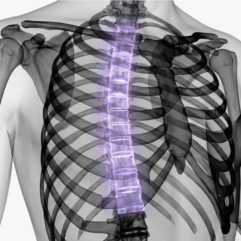
A thoracic spine MRI is an examination using magnetic resonance imaging (MRI) of the middle section of the spine, known as the thoracic spine. This area consists of twelve thoracic vertebrae and extends from the base of the neck to the upper part of the lumbar spine. The thoracic spine surrounds and protects a portion of the spinal cord and is connected to the rib cage via the ribs. An MRI of the thoracic spine provides detailed images of structures such as vertebrae, intervertebral discs, the spinal cord, the spinal canal, and surrounding soft tissues. The examination is particularly useful for assessing structures that are not clearly visible on standard X-rays. The MRI scanner uses a strong magnetic field and radio waves; therefore, it does not involve ionizing radiation.A thoracic spine MRI may be indicated for persistent or unexplained mid-back pain, but especially when symptoms suggest involvement of the discs, spinal cord, vertebrae, or other deep-seated structures. Depending on the clinical question, the examination may also be used to supplement findings from previous X-rays or CT scans.
What can be seen with a thoracic spine MRI?
An MRI scan of the thoracic spine provides detailed information about various structures within and around the spine. The radiologist can assess, among other things:
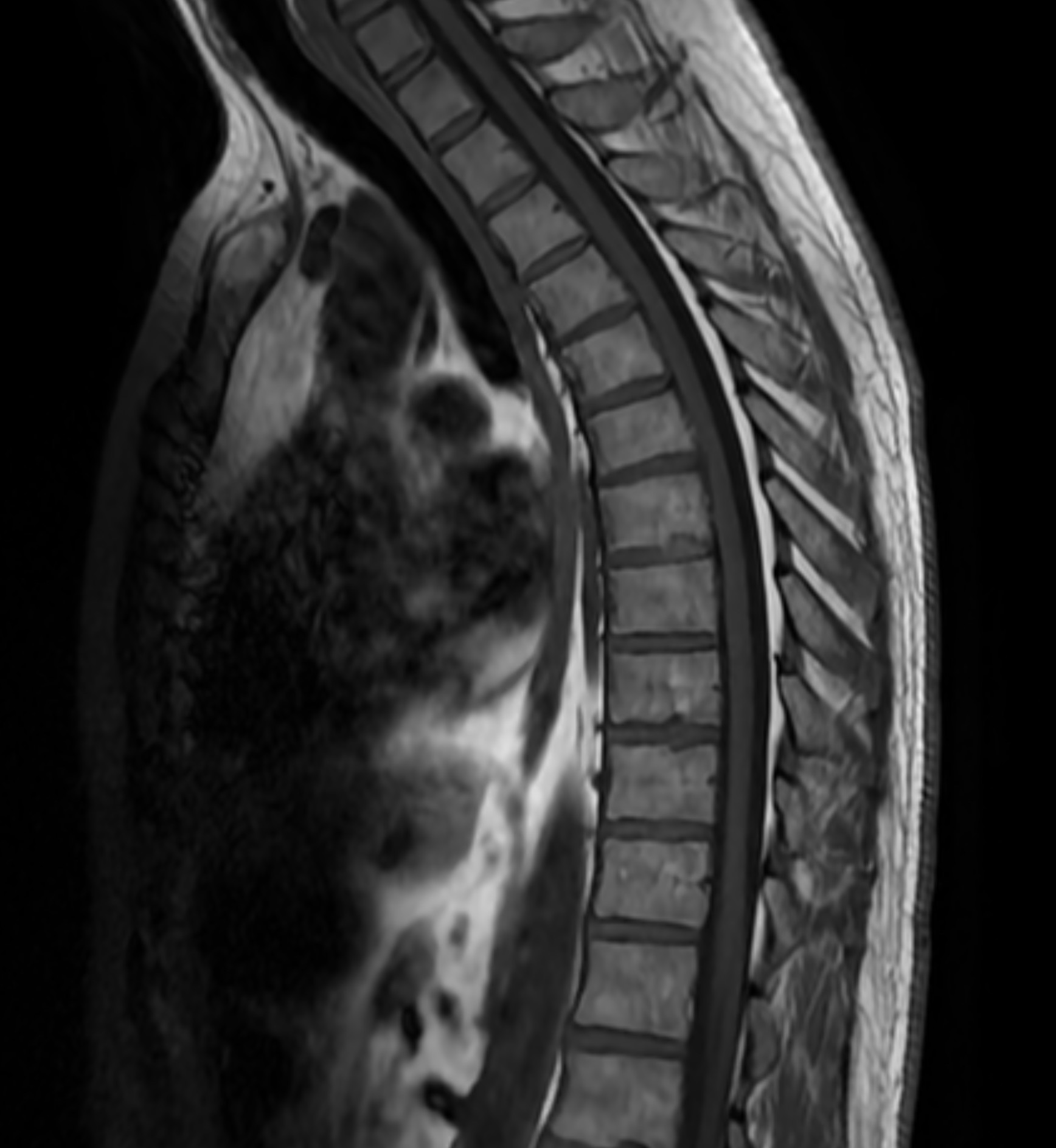
MRI image of the thoracic spine showing the thoracic vertebrae, intervertebral discs, spinal canal, and spinal cord. MRI provides detailed images of spinal structures and can be used to assess, for example, changes in the discs or vertebrae and any impact on the spinal cord.
- Thoracic vertebrae and vertebral bodies.
- Intervertebral discs and signs of disc degeneration.
- Disc bulging or herniation.
- The spinal cord and the space surrounding it.
- The spinal canal and any narrowing.
- Changes in the bone marrow.
- Inflammatory or infectious changes.
- Suspected tumors or metastases.
- Surrounding ligaments and soft tissues.
When might an MRI of the thoracic spine be appropriate?
An MRI of the thoracic spine may be appropriate when symptoms are persistent or recurrent, or when accompanied by signs suggesting involvement of the nervous system or deeper spinal structures. The most suitable examination depends on the symptoms, medical history, and the specific clinical question being addressed.
- Prolonged or unexplained pain in the mid-back or between the shoulder blades.
- Pain radiating around the chest or along the ribs.
- Numbness, tingling, or altered sensation in the torso.
- Weakness, sensory changes, or other neurological symptoms affecting the legs.
- Suspected disc bulge or herniated disc in the thoracic spine.
- Suspected spinal cord involvement.
- Suspected vertebral compression fracture or other vertebral abnormality.
- Suspected inflammation or infection of the spine or discs.
- Evaluation in cases of known cancer or suspected spinal tumor.
- Follow-up of a previously identified abnormality or post-operative assessment of the thoracic spine.
Thoracic spine MRI for herniated discs and disc bulges
Herniated discs are significantly more common in the cervical and lumbar spine than in the thoracic spine, but they can also occur between the thoracic vertebrae. A disc may bulge or rupture, thereby affecting the space within the spinal canal. MRI is the imaging modality that clearly visualizes the intervertebral discs and their relationship to the spinal cord. The examination can therefore be used to assess whether a disc abnormality is causing compression or other impact on nerve tissue.
Thoracic spine MRI for spinal cord involvement
A key reason for performing an MRI of the thoracic spine is suspected involvement of the spinal cord. Since the spinal cord runs through the spinal canal, it can be affected by conditions such as disc abnormalities, narrowing (stenosis), tumors, or other pathological processes.
Symptoms of spinal cord involvement can include altered sensation, numbness, leg weakness, walking difficulties, or impaired coordination. MRI provides detailed images of both the spinal cord and surrounding structures, making it a key diagnostic tool for this type of clinical question.Vertebral compression and changes in the thoracic vertebrae
Vertebral compression means that a vertebra has collapsed. This can occur, for example, due to osteoporosis, following trauma, or in association with other skeletal changes. The thoracic spine is a common site for vertebral compressions. MRI can provide information on the shape of the vertebrae and the bone marrow, and in some cases, help determine whether a change is recent or longstanding. If a fracture is suspected, X-ray or computed tomography (CT) may also be appropriate, depending on the situation.
Inflammation and infection in the thoracic spine
>MRI is sensitive to changes in bone marrow and soft tissues and can therefore be used when inflammation or infection of the spine is suspected. Examples include infection of a vertebra or an intervertebral disc—conditions known as spondylitis and discitis, respectively. If infection, a tumor, or certain inflammatory conditions are suspected, a contrast agent may be required to provide additional diagnostic information. This is determined based on the clinical question and the examination protocol.
Thoracic spine MRI for tumors or metastases
In cases of known cancer or suspected tumors, MRI can be used to examine the thoracic vertebrae, bone marrow, spinal cord, and tissues surrounding the spine. The examination helps map the location and extent of an abnormality and determine whether the spinal canal or spinal cord is affected. Diagnostic imaging is part of the medical workup, and MRI findings must always be evaluated in conjunction with other examinations and clinical data.
Thoracic spine MRI vs. X-ray – what is the difference?
Conventional X-ray is primarily useful for visualizing the shape of the skeleton and can be used, for instance, to assess certain fractures or deformities. MRI, on the other hand, provides significantly more detailed information about discs, the spinal cord, bone marrow, and soft tissues. The most suitable method therefore depends on what is being examined. MRI is often particularly valuable in cases involving neurological symptoms where involvement of the spinal cord or nerve structures is suspected.
When should you seek emergency care?
Back pain accompanied by a new onset of muscle weakness, difficulty walking, extensive loss of sensation, or problems controlling urine or bowel movements can be signs of serious nerve or spinal cord involvement. If you experience such symptoms, you should seek emergency medical care rather than waiting for a scheduled MRI scan.
Book an MRI of the thoracic spine
An MRI of the thoracic spine is an imaging examination that provides detailed images of the thoracic vertebrae, discs, spinal cord, and spinal canal. The examination can be valuable when investigating persistent symptoms or when an abnormality affecting the deeper structures of the spine is suspected. Upon booking, the medical referral is processed, and the examination is performed at an affiliated radiology clinic. The images are reviewed by a specialist radiologist, and you will receive a medical report containing the results after the examination.