
Chest MRI (Thorax) – MRI examination of the chest wall, soft tissues, and deep structures
Chest MRI (also known as thoracic MRI) is an imaging procedure that provides detailed images of the soft tissues and deeper anatomical structures of the chest. The examination can be used to assess, among other things, the chest wall, muscles, soft tissues near the ribs, the mediastinum, the pleura (lining of the lungs), lymph node regions, and other tissues within the thorax.
MRI is particularly effective at distinguishing between different types of soft tissue; it is therefore valuable for detailed characterization of an abnormality—such as a lump, swelling, suspected inflammation, or a lesion in the chest wall. MRI does not use ionizing radiation and can generate images of the chest in multiple planes.
Unlike computed tomography (CT)—which is often the primary method for examining lung tissue itself—thoracic MRI is primarily used when the clinical question concerns soft tissues, the chest wall, the mediastinum, or other structures where the high soft-tissue contrast provided by MRI offers an advantage.
What can be seen on a chest MRI?
During a chest MRI examination, the radiologist can evaluate a range of structures within the thoracic region. The specific areas examined and the imaging sequences used are tailored to the clinical question at hand.
- Chest wall muscles and soft tissues.
- Abnormalities in or adjacent to the ribs and sternum.
- Mediastinum – the area between the lungs.
- Lymph nodes and soft-tissue structures in the thorax.
- Pleura and tissues adjacent to the lungs.
- Lumps, cystic lesions, and other soft-tissue abnormalities.
- Inflammatory changes in muscles or soft tissues.
- Extent of a previously identified abnormality in the chest.
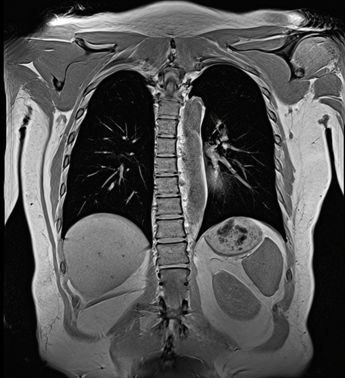
Coronal MRI image of the chest (thorax) showing the lungs, chest wall, spine, mediastinum, and parts of the upper abdomen. The imaging plane provides a clear overview of the chest anatomy from a frontal perspective.
When might a chest MRI be indicated?
A chest MRI may be indicated when symptoms or findings from previous examinations suggest a need for a more detailed assessment of the chest's soft tissues or deeper structures. The examination can also be used as a supplement to procedures such as X-ray, ultrasound, or CT scanning.
- Lump or swelling in the chest wall or the soft tissues of the chest.
- Persistent localized pain in the chest wall with no clear explanation.
- Suspected tumor or other soft-tissue abnormality.
- Abnormality in the mediastinum or lymph node region requiring further characterization.
- Suspected inflammation in muscles, the chest wall, or other soft tissues.
- Abnormalities in or around the pleura.
- Follow-up of a previously identified abnormality.
- Assessment following previous surgery or other treatment in the thoracic region.
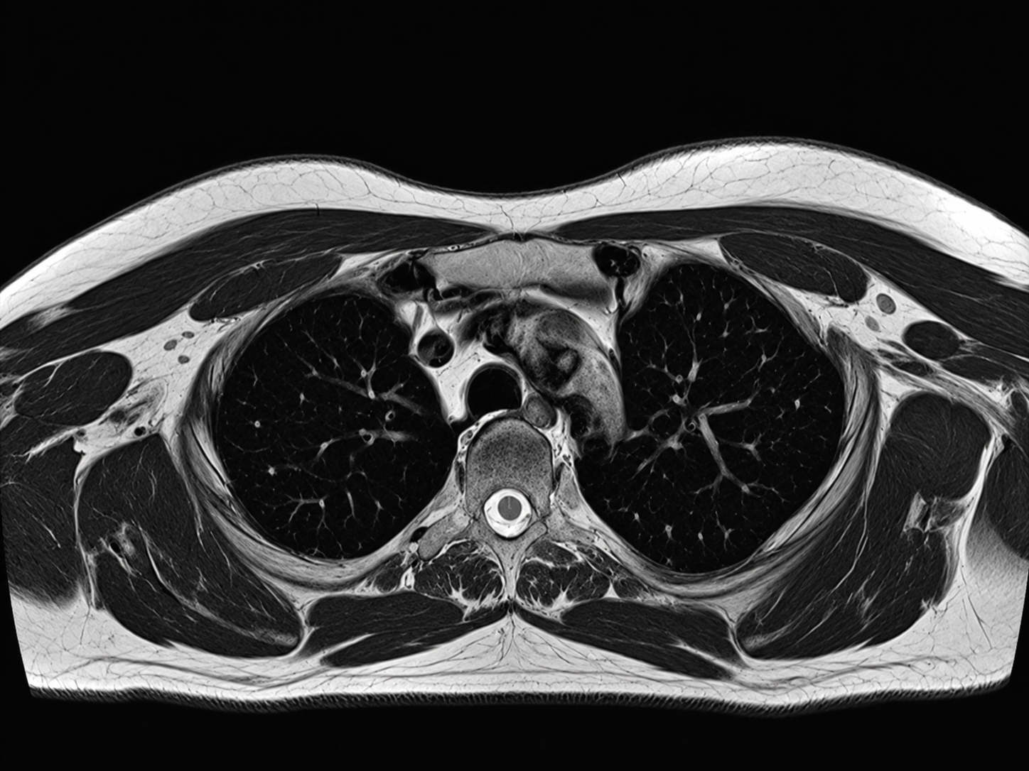
Axial MRI image of the chest (thorax) showing the lungs, mediastinum, chest wall, spine, and surrounding soft tissues. The imaging plane shows the chest in cross-section, providing a detailed overview of the thoracic structures.
Chest MRI for lumps, swelling, or localized pain
A lump or swelling in the chest area can originate from various tissues, such as muscle, connective tissue, adipose tissue, or chest wall structures. MRI provides detailed information regarding the size and location of the abnormality and its relationship to surrounding tissues. In cases of persistent or localized pain, the examination can sometimes help identify abnormalities in muscles and other soft tissues. However, the most suitable imaging method depends on the symptoms and the specific location of the issue within the chest.
Chest MRI for suspected tumors and soft-tissue abnormalities
MRI offers excellent soft-tissue contrast resolution and is therefore frequently used to characterize and map soft-tissue abnormalities. For instance, MRI can provide information on the extent of an abnormality and its relationship to muscles, the chest wall, and adjacent structures. When a tumor is suspected, diagnostic imaging forms part of the medical workup. An MRI scan alone cannot definitively identify the specific type of abnormality, so further examinations or tissue sampling may be required.
MRI of the mediastinum and lymph node regions
The mediastinum is the central compartment of the chest located between the lungs; it contains structures such as major blood vessels, the trachea, the esophagus, lymph nodes, and other soft tissues. In selected cases, MRI is used to assess abnormalities in this area in greater detail and to evaluate their relationship to surrounding tissues. Enlarged lymph nodes can have a wide range of causes.
MRI can show their location and extent, but diagnostic imaging findings must always be evaluated alongside other medical findings to determine the cause.Thoracic MRI vs. Thoracic CT – what is the difference?
Both MRI and CT scans can be used to examine the chest, but the methods have different strengths. A thoracic CT scan is very fast and particularly effective for assessing lung tissue, airways, and many acute conditions. Thoracic MRI, on the other hand, provides highly detailed information about soft tissues and is therefore primarily used for specific clinical questions where this tissue contrast is important.
Furthermore, MRI does not use ionizing radiation. The most suitable examination is determined by the specific structure or disease being investigated.
Examination of the heart and major blood vessels
The heart and major blood vessels are located in the chest, but a detailed examination of these structures often requires specialized imaging protocols. For questions primarily concerning the heart, a dedicated cardiac MRI might be more appropriate, whereas vascular issues may require MR angiography or other forms of diagnostic imaging.
Therefore, a general thoracic MRI should not be equated with a cardiac examination or a comprehensive vascular assessment.
Is a contrast agent needed for a thoracic MRI?
In some cases, an intravenous contrast agent may be used to provide additional information regarding, for example, a soft-tissue abnormality or an inflammatory process. The need for contrast agents is determined based on the clinical indication and the examination protocol.
Chest MRI does not use X-rays
The MRI scanner creates images using a strong magnetic field and radio waves; therefore, it does not use ionizing X-rays. The examination is non-invasive and allows for imaging of the same area from multiple directions.
Book a Chest MRI
A chest MRI is an imaging examination for those requiring a detailed assessment of the chest wall, soft tissues, or other structures within the thoracic region. Your medical referral is processed in conjunction with your booking, and the examination is performed at an affiliated radiology clinic. The images are reviewed by a specialist radiologist, and following the examination, you will receive a medical report containing the results.