
MR Kidneys – Magnetic resonance imaging examination for kidney problems, cysts or suspected tumors
Have you had blood tests that showed abnormal kidney values, or are you experiencing symptoms such as fatigue, back pain, swelling or recurrent urinary tract problems? Then an MRI examination of the kidneys can be an important next step. MRI kidneys is a gentle, radiation-free and very accurate method for detecting cysts, tumors, inflammation or other changes in the kidneys that may explain your symptoms.
The examination provides high-resolution images of the structure of the kidneys and surrounding tissue and is used both in cases of suspected disease and as a follow-up of previous findings. Since MRI does not use X-rays, it is also suitable for people with impaired kidney function or hypersensitivity to contrast agents.
When is an MRI of the kidneys recommended?
MRI of the kidneys is used as a safe and gentle alternative when there is a need to evaluate morphological or functional changes – both in acute and chronic conditions.
- Blood in the urine (hematuria) without a clear cause
- Suspected tumor, cyst or change in the kidney tissue
- Follow-up of known kidney changes
- Kidney-related pain or swelling in the flanks
- Urinary outflow obstruction or hydronephrosis
- Congenital malformations or anatomical variations
- Investigation in chronic kidney disease, with the need for detailed imaging diagnostics
MRI is often used when the following conditions in the kidneys are suspected
- Kidney cancer or tumor in the renal pelvis
- Simple or complex kidney cysts
- Hydronephrosis (fluid accumulation due to outflow obstruction)
- Urinary tract stones (stones) in the upper urinary tract
- Pyelonephritis or other inflammation of the kidney
- Changes after surgery or transplantation
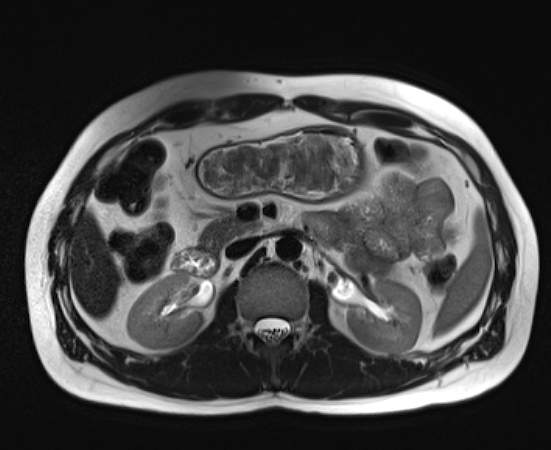
Axial MRI image of the kidneys and upper urinary tract showing the anatomy of the kidneys, the renal pelvis and surrounding abdominal organs.
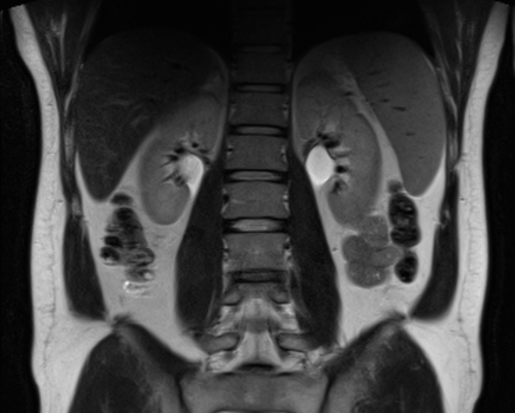
MRI image of the kidneys showing the anatomy of the kidneys and surrounding tissues in detail. The examination can be used to detect and assess various changes in the kidneys.
Can kidney stones be seen on MRI kidneys?
MRI is not the first-line method for detecting kidney stones. Computed tomography (X-ray) is mainly used to evaluate suspected or known kidney stones, which provides a clear image and can reliably detect possible kidney stones. Kidney stone attacks are caused when kidney stones become lodged in the ureter and block the flow of urine from the kidney, which builds up high pressure that causes intense pain that is usually felt over the flank or forward in the abdomen and groin. If you suspect that you have ongoing kidney stone pain, this examination is not recommended, but you should contact your nearest emergency department for care.
Book MRI Kidneys – A radiation-free and safe examination
An MRI examination of the kidneys takes approximately 30–40 minutes and is performed in the supine position. The examination is completely painless and does not involve exposure to radiation. You do not need your own referral, we issue it directly in connection with your booking. The results are analyzed by a specialist in radiology and you will receive a written report within a few days.