
MRI Wrist

5 195 kr

MRI Wrist

Magnetic resonance imaging

Information

Medical comment included

With us, you will always receive a doctor’s opinion from a licensed physician when you order a medical check-up that includes a medical opinion. The doctor’s opinion includes an overall assessment of your results from laboratory analysis. The opinion is individual and may vary depending on the specific tests your medical check-up includes.

What can I expect in the medical report?

The medical opinion will summarize the results of your test results to give you an assessment of your health including any abnormalities outside the reference ranges. In case of abnormalities or need, your doctor may also contact you for a free conversation to explain and talk about your results. Remember that a health check always includes specific test results based on your health and your personal circumstances. Values outside the reference range should therefore always be discussed with a doctor before any decisions regarding possible treatment are made.

What does the doctor look at during a health check?

When assessing a blood test or health check, your doctor will look at several different parameters to evaluate your health and identify any abnormalities. Below are some common aspects your doctor may examine:

Blood counts: Your doctor will check red, white and platelet counts to identify any signs of anaemia, infection or bleeding.

Nutrients and biochemical markers: Includes, for example, analysis and monitoring of blood sugar (glucose) levels, electrolyte levels (e.g. sodium, potassium), liver function tests (e.g. liver enzymes), kidney function tests (e.g. creatinine, urea) and lipid profile (e.g. cholesterol, triglycerides). The results of the tests give your doctor a better idea of your organ function, blood sugar level, cholesterol conditions and other important health aspects.

Inflammatory markers: Your doctor may measure health markers such as C-reactive protein (CRP) or red blood cell decrease rate (SR) to evaluate inflammatory conditions or confirm an active infection.

Hormone levels: Your doctor may monitor your hormone levels to assess hormonal imbalances or diseases, such as thyroid hormones (TSH, T3, T4), sex hormones or other specific hormones such as testosterone levels.

Specific tests: Depending on your goal of your health check or medical history, your doctor may look into specific tests to investigate conditions or diseases, such as diabetes, vitamin deficiency or autoimmune diseases.

In conclusion, the doctor weighs all test results based on the health check to assess your overall health, detect any abnormalities and identify possible underlying medical conditions.

What happens if I have abnormal test results?

If your blood count is abnormal, this will be addressed in your personal medical opinion. Depending on the nature and extent of the abnormality, your doctor may also contact you directly through the test result service for further guidance. Various measures and advice will be provided based on the specific abnormalities observed along with your medical history.

Call included

We always offer a phone consultation with a licensed physician before a referral for an MRI or ultrasound is issued. This allows us to ask the right questions and ensure that you receive the most appropriate and accurate examination for your needs. After the examination, you will receive both the radiologist’s report and a medical opinion, and we remain available to support you throughout the entire process. Follow-up consultations can be booked if desired. Once you have activated the referral, one of our licensed physicians will call you.


Any questions? Call us for advice at 08–14 24 49.

5 195 kr
  • Magnetic camera examination of the wrist without referral
  • Recommended for wrist pain or suspected ligament injury
  • Detailed MRI examination of tendons, joints and nerves
  • Quick booking and response – painless examination at Unilabs
Read more

Add-ons

Affiliate Icon

Here it is empty.

Try removing filters or reloading.
Reload
Lågt pris
Lowest price on the market
Ingen strålning
No radiation
Ingen strålning
Referral within 1 business day

MRI wrist provides detailed images of the skeleton, ligaments and tendons

The wrist is an anatomically complex joint that consists of eight small carpal bones together with the radius and ulna of the forearm. Between the bones are several small joints and ligaments that together give the wrist stability while allowing a large range of motion. A large number of tendons, nerves and blood vessels also pass through the area.

Pain or impaired function in the wrist can therefore be due to many different structures and it is not always possible to determine the cause solely through symptoms and clinical examination. Magnetic resonance imaging, MRI, can provide detailed images of both the skeleton and soft tissues and is particularly valuable for assessing ligaments, TFCC, tendons, bone marrow, cartilage and other soft tissue structures.

MRI wrist is often used when the symptoms have persisted for a long time, when an injury has not healed as expected or when previous X-ray examinations have not been able to explain the symptoms. The examination uses magnetic fields and radio waves and does not involve ionizing radiation.

When can an MRI of the wrist be relevant?

Which imaging examination is most appropriate depends on the symptoms and the suspected injury. In acute trauma or long-term wrist pain, plain X-rays are often used as a first step to assess the skeleton. If the X-ray is normal or does not provide sufficient information, MRI can be an important next step.

MRI of the wrist may be relevant in, among other things:

  • persistent or recurrent wrist pain where the cause has not been determined.
  • pain after a fall, twist or other injury when the X-ray does not explain the symptoms.
  • suspected injury to ligaments or TFCC.
  • swelling, stiffness or pain that may be due to inflammation.
  • clicks, locking or a feeling of instability in the wrist.
  • suspected damage to tendons or tendon sheaths.
  • suspected fracture or stress injury that is not clearly visible on a regular X-ray.
  • suspected ganglion or other soft tissue changes.

MRI can also be used before treatment or surgery when there is a need to map the extent of an injury and its relationship to surrounding structures.

What can an MRI of the wrist show?

An MRI provides images in multiple planes and has high contrast between different soft tissues. This makes it possible to examine several of the wrist's small anatomical structures that can be difficult to assess with standard X-rays.

The examination can provide information about, among other things:

  • the bones and bone marrow of the wrist.
  • the scapholunate and lunotriquetral ligaments.
  • TFCC, triangular fibrocartilaginous complex.
  • tendons and tendon sheaths.
  • articular cartilage and joint surfaces.
  • synovial fluid and inflammatory changes.
  • ganglion and other soft tissue changes.
  • signs of hidden fracture or stress reaction in the skeleton.

Exactly what can be assessed depends on the question and which MRI protocol is used. In some specific questions, other examinations, such as ultrasound, computed tomography or MRI arthrography, may be more appropriate.
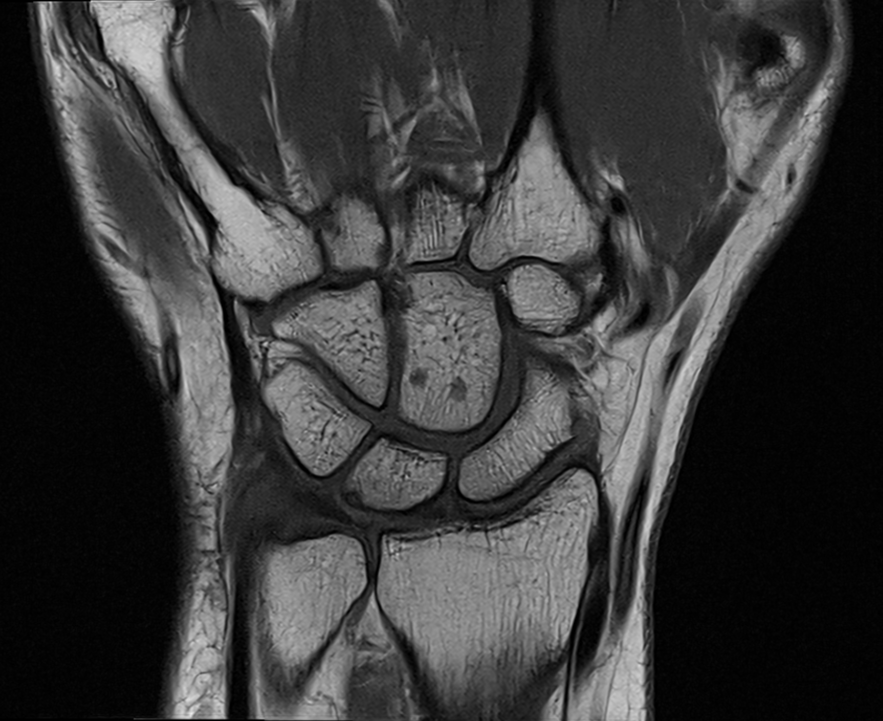
MRI image of the wrist

The image shows an example of an MRI image of the wrist. In the same examination, the radiologist can assess the small carpal bones, the lower part of the radius and ulna, as well as several of the wrist's ligaments and soft tissues. MRI can also show changes within the bone marrow that are not always visible on regular X-rays.

TFCC injury

TFCC, triangular fibrocartilage complex, is a complex of cartilage and ligament structures on the little finger side of the wrist. The structure contributes to the stability between the bones of the forearm and the wrist and also acts as a shock absorber during loading. TFCC can be damaged by, for example, falling on an outstretched hand, heavy rotation or repeated loading. Common symptoms are pain on the little finger side of the wrist, clicking, weakness when gripping and discomfort when twisting the forearm. MRI can be used to assess TFCC, but in certain cases, MRI arthrography or other complementary examination can provide additional information.

Ligament injuries and instability

The small carpal bones are held together by several ligaments. Two particularly important structures are the scapholunate ligament between the scaphoid and lunate and the lunotriquetral ligament between the lunate and triquetrum. Damage to these ligaments can lead to pain, clicking and instability and, in the long term, contribute to altered load and the development of osteoarthritis. MRI can show the ligaments themselves and provide information about the surrounding soft tissues and bone marrow, but the diagnosis may sometimes need to be supplemented depending on the type of injury.

Scaphoid fracture and hidden fractures

The scaphoid, or scaphoid, is one of the small carpal bones of the wrist and can be injured, for example, when falling on an outstretched hand. A scaphoid fracture can sometimes be difficult to see on the first X-ray examination. If clinical suspicion remains, MRI can be used to identify a fracture that is not visible on a regular X-ray.

MRI is sensitive to changes in the bone marrow and can therefore show both fractures and bone marrow edema. In the case of an already known navicular fracture, MRI or computed tomography can also be used for specific issues such as failure to heal or effects on the blood supply to the bone.

Tendons and tendon sheaths

A large number of flexor and extensor tendons pass through the wrist and enable movements in the hand, fingers and thumb. MRI can show changes in the tendons and tendon sheaths, such as inflammation, fluid accumulation, degenerative changes and certain tendon injuries. In de Quervain's disease, the tendon sheaths on the thumb side of the wrist are affected and typical symptoms are pain in the thumb and wrist, especially when gripping and moving. If there is a clear suspicion of superficial tendon problems, ultrasound can also be a very useful examination method.

Arthritis and inflammatory joint diseases

MRI can show changes in joint surfaces, bone marrow, synovial membrane and soft tissues and can therefore provide information in osteoarthritis and inflammatory joint diseases. In inflammatory arthritis, MRI can show synovitis, tenosynovitis, bone marrow edema and erosions. In contrast, regular X-rays are often used as an initial examination in suspected osteoarthritis or chronic joint disease. MRI is used primarily when more detailed information is needed or when symptoms and previous examinations do not match.

Ganglion in the wrist

A ganglion is a fluid-filled lesion that often occurs in connection with a joint or tendon sheath. Ganglions are common around the wrist and can be completely asymptomatic but can also cause pain, pressure or discomfort during movement. MRI can show the size and location of the lesion and its relationship to joints, ligaments, tendons and other surrounding structures. In many cases, a superficial ganglion can also be assessed with ultrasound.

Carpal Tunnel Syndrome and Nerve Impairment

Carpal tunnel syndrome is caused by pressure on the median nerve as it passes through the carpal tunnel in the wrist. Common symptoms include numbness, tingling, and sometimes pain in the thumb, index finger, middle finger, and parts of the ring finger.

MRI can show the median nerve and structures in the carpal tunnel but is not routinely used as a first-line examination for typical carpal tunnel syndrome. The diagnosis is based primarily on symptoms and clinical examination and can be supplemented with neurophysiological examination if necessary. MRI may be relevant when symptoms are atypical or when a structural cause of nerve damage needs to be ruled out.

How is a wrist MRI performed?

During the examination, the wrist is placed in a special coil that makes it possible to take detailed images of the relatively small anatomical area. Images and sequences are taken in several different planes so that the radiologist can assess the skeleton, joints, ligaments, tendons and soft tissues from different angles. The image below shows a different section through the wrist than the previous MRI image and illustrates how the same anatomical area can be examined from several directions for a more complete assessment.
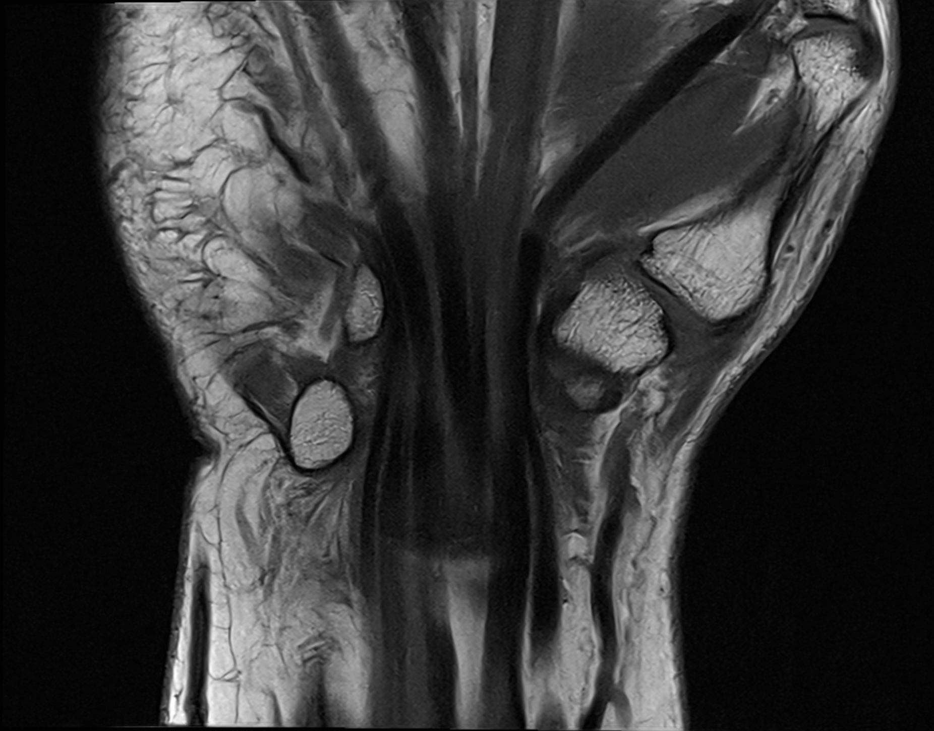
MR image of the wrist in a different image plane showing the carpal bones and surrounding soft tissues.

The examination usually takes about 20 to 30 minutes. MRI does not use any X-ray radiation and the examination itself is painless, but it may be uncomfortable to lie still for a long time. In certain specific questions, contrast media may be needed, but many examinations of the wrist are performed without intravenous contrast media.

Book MRI wrist

After ordering, you will receive a call from one of our doctors who will review your medical history and your symptoms. A referral will then be sent directly to the X-ray clinic. MRI of the wrist may be particularly relevant in cases of long-term or difficult-to-assess wrist pain, suspected ligament or TFCC injury, tendon injury or when previous X-rays have not been able to explain the symptoms. The results are assessed by a radiologist and can then be used as a basis for further medical assessment and possible treatment. If you have problems primarily further out in the hand or fingers, an examination of the hand may be more relevant than an MRI of the wrist. Which area should be examined is determined by where the symptoms are located and which structure is suspected to be affected.

The most mobile extremities of the body are the arms, which consist of several joints, muscles and bones that together enable both power and precision. The upper arm runs down from the shoulder to the elbow, where the humerus meets the two bones of the forearm – the radius and ulna. The wrist and hand are extremely mobile and have fine motor skills, which makes it possible to grasp, write, point and perform other complex movements.

The muscles of the arms are divided into flexors (which bend) and extensors (which stretch), and nerves from the cervical spine control both sensation and movement. Blood vessels and tendons run along the entire arm, supplying each part with oxygen and nutrients. The nails, especially on the fingers, serve as protection and tools for sensation and precision. In summary, our arms are central to how we interact with our surroundings.

WRL
Wrist
The wrist is a complex joint that connects the forearm to the hand and allows for movement in multiple directions. It is important for fine motor skills and everyday movements.
Read more

After your order, we will send a referral for your MRI scan. Before that, you will have a brief consultation with a referring physician who will review your medical history and symptoms. After the consultation, your referral will be forwarded to the radiology clinic.

Magnetic Resonance Imaging (MRI) is a safe method for obtaining detailed images of the body's organs, tissues, and joints using a strong magnetic field and radio waves – completely without radiation.

Things to consider before the examination:

  • Clothing and items: You will be asked to remove clothing with metal, jewelry, watches, hearing aids, and other items that may be affected by the magnetic field.
  • Metal in the body: Inform the medical staff if you have a pacemaker, implants, metal clips, artificial joints, or other metal objects in your body.
  • Pregnancy: Notify the staff if you are pregnant or suspect that you might be.
  • Contrast agent: In some cases, a contrast agent is used to enhance image quality. You will be informed about any preparations or possible side effects if applicable.

After your MRI scan is completed, the images are reviewed by a specialist in radiology. The radiologist interprets the images based on the clinical question stated in your referral and prepares a medical report for the referring physician.

The referring physician from our team will then review the report and provide you with a written medical assessment and guidance.

If the examination reveals any abnormalities or findings, we will ensure that you are either referred to an appropriate healthcare provider or receive advice on suitable follow-up – all of this is included in the price.

Find the nearest test location

How does it work?

Order an MRI scan – four easy steps

Step 1

Order MRI

Select the MRI scan you wish to perform and add to your cart, for example lumbar spine, abdomen or full body. Complete your order and select a payment option, we always apply an open purchase on MRI for at least 30 days.

Step 2

Activate the referral

After payment, you’ll receive an email with instructions. Log in to enter your details and activate your referral. A physician will contact you to confirm your booking and discuss your preferences.

Step 3

Complete your MRI

You will be called via Kivra or mail within 7–10 working days, visit the clinic you have been called to at the appointed time. Contact the clinic directly for possible rescheduling if the time does not suit you. Read more

Step 4

Results and statement

You will receive feedback on your X-rays within 5–7 working days. However, in some cases it may take a few extra days before all results are ready and reviewed by specialist doctors. Read more

Your test answers in zample™

– A smarter tool for understanding your health

Select area

See the full list below

Other MRI examinations


MRI Full body Plus

MRI Full body Plus

MRI + blood tests

21 000 kr

MRI Full Body Pro

MRI Full Body Pro

MRI + blood tests + ECG

23 900 kr

Prostate Check – MRI & Stockholm3

Prostate Check – MRI & Stockholm3

MRI Prostate + Stockholm3

10 599 kr


MRCP

MRCP

MRI of the biliary tract and pancreas

7 695 kr

MRI Abdomen/MRCP

MRI Abdomen/MRCP

MRI of the abdomen

6 995 kr

MRI Aorta

MRI Aorta

MRI

9 495 kr

MRI Brain

MRI Brain

Magnetic resonance imaging

6 795 kr

6 795 kr

9 195 kr

9 195 kr

MRI Ears

MRI Ears

Magnetic resonance imaging

5 595 kr

13 995 kr

MRI Full Body

MRI Full Body

Magnetic resonance imaging

19 995 kr

MRI Full body Plus

MRI Full body Plus

MRI + blood tests

21 000 kr

MRI Full Body Pro

MRI Full Body Pro

MRI + blood tests + ECG

23 900 kr

MRI Kidneys

MRI Kidneys

MRI scan of the kidneys

6 195 kr

MRI Pancreas

MRI Pancreas

Magnetic resonance imaging

6 195 kr

MRI Pelvis

MRI Pelvis

MRI scan

6 495 kr

MRI Pituitary gland

MRI Pituitary gland

Magnetic resonance imaging

4 495 kr

6 395 kr

MRI Prostate

MRI Prostate

Magnetic resonance imaging

7 295 kr

7 295 kr

MR Auditory pathway

MR Auditory pathway

MRI of the pore

4 995 kr

MR Liver

MR Liver

MRI examination of the liver

6 795 kr


MRI Ankle

MRI Ankle

Magnetic resonance imaging

4 495 kr

MRI Cervical spine

MRI Cervical spine

Magnetic resonance imaging

5 995 kr

MRI Chest

MRI Chest

Magnetic resonance imaging

6 795 kr

MRI Elbow

MRI Elbow

Magnetic resonance imaging

4 495 kr

MRI face

MRI face

Magnetic resonance imaging

5 795 kr

MRI Femur

MRI Femur

Magnetic resonance imaging

4 495 kr

MRI Foot

MRI Foot

Magnetic Resonance Imaging

5 195 kr

MRI Forearm

MRI Forearm

Magnetic Resonance Imaging

3 795 kr

13 995 kr

MRI Full Body

MRI Full Body

Magnetic resonance imaging

19 995 kr

MRI Full body Plus

MRI Full body Plus

MRI + blood tests

21 000 kr

MRI Full Body Pro

MRI Full Body Pro

MRI + blood tests + ECG

23 900 kr

MRI Hand

MRI Hand

Magnetic resonance imaging

5 195 kr

MRI Hip joint and pelvis

MRI Hip joint and pelvis

Magnetic resonance imaging

5 795 kr

MRI Knee

MRI Knee

Magnetic Resonance Imaging

5 395 kr

MRI Knee

MRI Knee

Magnetic Resonance Imaging

5 395 kr

MRI Lower Leg

MRI Lower Leg

Magnetic resonance imaging

4 495 kr

MRI Lumbar spine

MRI Lumbar spine

MRI of the lumbar spine

5 995 kr

MRI Orbita

MRI Orbita

Magnetic resonance imaging

5 395 kr

MRI Sacroiliac joints

MRI Sacroiliac joints

Magnetic Resonance Imaging

4 495 kr

MRI Shoulder

MRI Shoulder

Magnetic resonance imaging

4 495 kr

MRI Sinus

MRI Sinus

Magnetic Resonance Imaging

6 495 kr

MRI Skull base

MRI Skull base

Magnetic Resonance Imaging

4 495 kr

MRI Temporomandibular joints

MRI Temporomandibular joints

Magnetic resonance imaging

5 495 kr

MRI Thoracic spine

MRI Thoracic spine

Magnetic resonance imaging

4 995 kr

MRI throat

MRI throat

Magnetic resonance imaging of the throat

6 895 kr

MRI Upper arm

MRI Upper arm

Magnetic Resonance Imaging

4 495 kr

MRI Wrist

MRI Wrist

Magnetic resonance imaging

5 195 kr

MR Achilles tendon

MR Achilles tendon

Magnetic resonance imaging

4 195 kr

5 595 kr

With us you are always safe with our satisfied customer guarantee!

With us you are always safe with our satisfied customer guarantee!

We want to help people improve their health. That's why it's important that you feel safe with us. As a healthcare provider, safety is our top priority and we always strive to deliver the highest quality in our services. If for any reason you are not completely satisfied with your experience with us - do not hesitate to contact us. We listen and will do what we can to make you feel safe and satisfied. Our satisfied customer guarantee is an expression of our commitment to you and your health - where we want to take our responsibility when you use our services to access your health data and improve your health.