Quick version
A hepatic hemangioma is the most common benign tumor of the liver and is not cancer. It consists of blood-filled vascular spaces and is usually discovered by chance during an ultrasound, MRI or CT scan performed for other reasons.
Most hemangiomas are small, do not cause symptoms and do not affect liver function. Once the diagnosis has been confirmed with typical imaging findings, treatment or regular check-ups are usually not needed.
If the finding does not look completely typical, the doctor may want to perform additional imaging, for example with MRI or a contrast-enhanced CT scan. This is usually done to confirm that the lesion is indeed a hemangioma, not because something dangerous is already suspected.
Treatment is uncommon and is mainly relevant if the hemangioma is large, causes clear discomfort or leads to complications. Routine blood tests are often normal in people with hemangiomas, but liver function tests can still be valuable for detecting other causes of abnormal liver values or diffuse symptoms.
If you have been diagnosed with a small, typical hemangioma and feel well, the situation is usually calm. Seek medical attention if you experience increasing pain under the right rib cage, jaundice, fever or unexplained weight loss, or if the liver finding is discovered at the same time as you have known liver disease or a previous cancer diagnosis.
A liver finding often comes as a surprise. Someone may have an ultrasound because of gallbladder problems, a health check may show slightly elevated liver enzymes, or a CT scan may be performed for completely different reasons – and suddenly the report says that there is a hemangioma in the liver. For many people, the word “tumor” causes concern, even though this particular finding usually has a benign course.
What is a hemangioma in the liver?
A hemangioma in the liver, also called a liver hemangioma or hepatic hemangioma, is the most common benign tumor in the liver. It is not cancer, but a vascular lesion consisting of densely packed, blood-filled vascular spaces in the liver tissue. It is usually discovered by chance when someone is examined for another reason.
Many liver hemangiomas are small and never cause any symptoms. They can be present for years without affecting liver function or general health. Most people therefore do not need treatment or frequent check-ups once the diagnosis has been confirmed with typical imaging findings.
Hemangiomas are seen more often in women than in men, and they are usually discovered in adulthood. The reported prevalence varies between studies, partly depending on the examination method used and the population examined.
For those reading their radiology report at home, it is easy to get caught up in words such as “lesion”, “change” or “tumor”. In medical language, these words do not automatically mean that something is dangerous. In the case of hemangiomas, the key question is often not whether the lesion is aggressive, but whether the imaging pattern truly fits with a benign vascular lesion.
Common symptoms – and why many people do not feel anything at all
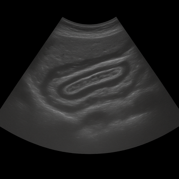
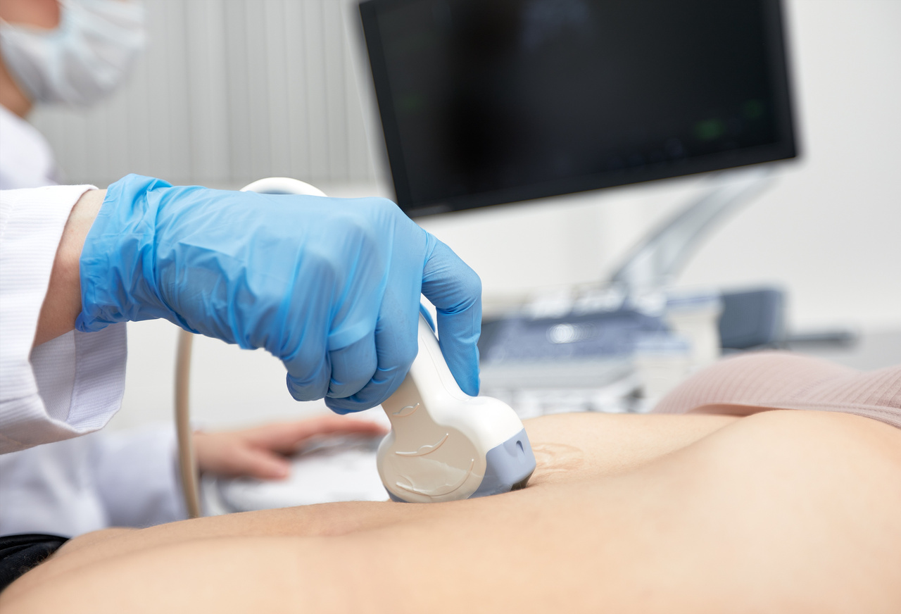
Most people with liver hemangiomas have no symptoms at all. The finding is often made during an abdominal ultrasound, MRI or CT scan ordered for other reasons, such as abdominal pain, kidney stones or investigation of the gallbladder.
When symptoms occur, they are usually related to larger lesions. In such cases, you may feel pressure or discomfort under the right rib cage, early satiety or diffuse upper abdominal pain. However, the symptoms are non-specific, which means that the same symptoms are also common in many other conditions, such as dyspepsia, gallstone disease or functional gastrointestinal problems.
A concrete example is a person being examined for recurrent bloating after meals, where an ultrasound finds a 2 cm hemangioma. In such a situation, the hemangioma is often just an incidental finding, while the symptoms have another explanation. Therefore, it is important not to attribute all abdominal complaints to a liver finding without making a reasonable overall medical assessment.
Very large hemangiomas can, in rare cases, cause more obvious problems by pressing on nearby structures. Rare complications do exist, but they are unusual and are seen primarily in large lesions or specific clinical situations.
How is the diagnosis made when a hemangioma in the liver is discovered?
The diagnosis is primarily based on imaging. A small hemangioma in the liver can sometimes already be recognized on ultrasound if it has a typical appearance, for example as a well-defined and brighter lesion in an otherwise healthy liver. In small and typical findings, this can sometimes be sufficient for diagnosis.
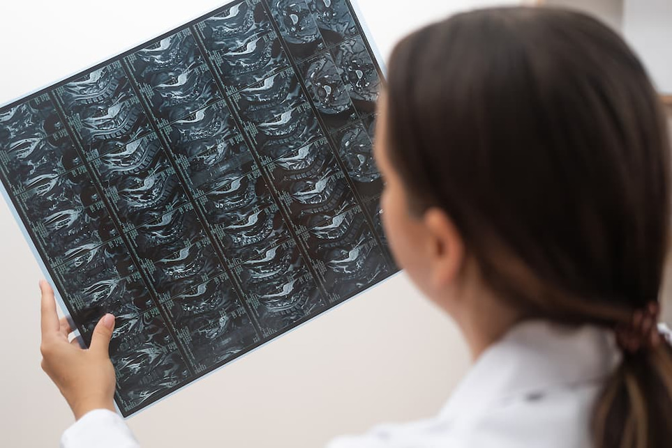
If the finding is not completely typical, it is often followed up with an MRI or contrast-enhanced CT scan. The radiologist then looks for typical imaging patterns that suggest a hemangioma and help distinguish it from other focal liver lesions.
MRI is particularly valuable when diagnostic certainty needs to be increased. This applies, for example, if the lesion is larger, if there is another concurrent liver disease or if the patient has a known cancer, where extra care is needed to distinguish benign findings from metastases.
A biopsy is not normally needed when a hemangioma is suspected. On the contrary, guidelines advise against needle biopsy if the imaging findings are typical, since the diagnosis can usually be made without a tissue sample and because vascular lesions may theoretically involve a risk of bleeding.
A common question from patients in this situation is: “If it is benign, why does the doctor want to take more images or investigate further?” The answer is that additional examinations are often done to confirm that the finding is indeed a hemangioma, not because the doctor already believes that it is something dangerous. The extra check is about precision, not panic.
When do liver hemangiomas need to be treated or followed up?
In most cases, no treatment is needed at all. When a liver hemangioma has a typical appearance on imaging and the person has no symptoms, routine follow-up is usually not recommended. This is especially true for small and stable lesions in people without other liver disease.
Treatment only becomes relevant when the hemangioma causes clear discomfort or complications. Examples may include pronounced pain where other explanations have been ruled out, pressure on organs or blood vessels, bleeding within the lesion or very unusual coagulation disorders such as consumption coagulopathy. In such cases, surgery or another intervention may be considered after specialist assessment.
Large hemangiomas are sometimes called “giant hemangiomas”, but there is no completely uniform size threshold in the literature. Five or ten centimetres are often used as practical cut-off values in studies and clinical reasoning. However, size alone does not determine everything – what matters is the combination of symptoms, growth, location and how certain the diagnosis is.
In pregnant women or people using estrogen therapy, it is sometimes discussed whether hormones can affect the size of a hemangioma. The state of knowledge is not entirely clear, but modern guidelines emphasize that pregnancy and birth control pills do not automatically mean that a typical hemangioma must be monitored more closely or treated. The assessment is individualized if the lesion is large or if symptoms occur.
When should you seek medical care and when can blood tests be valuable?
A liver hemangioma rarely shows up in regular blood tests. Liver tests such as ALT, AST, ALP, GGT and bilirubin can be completely normal, since liver function itself is usually not affected. If the tests are abnormal, it is often necessary to think more broadly and investigate other causes, such as fatty liver disease, alcohol exposure, medication effects, viral hepatitis or biliary tract disease. This makes blood tests relevant even when the imaging finding itself appears benign.
You should seek medical assessment if you experience new or increasing pain under the right rib cage, unexplained weight loss, fever, pronounced fatigue, jaundice or if a liver finding is discovered at the same time as you have known liver disease or previous cancer. In this case, the investigation is not only about the liver hemangioma, but about understanding the whole context.
If you have already been told that you have a small, typical hemangioma and are otherwise well, the situation is usually calm. In this case, it is often more valuable to follow your general health markers than to get stuck on the actual wording in the radiology report. In practice, this can mean checking liver values, blood sugar, blood lipids and other tests that reflect the broader health picture.
A hemangioma in the liver reminds us of an important aspect of modern medicine: the better we become at examining the body, the more often we find incidental findings that do not need to be treated, but that do need to be interpreted correctly. Real reassurance rarely comes from “looking more” endlessly, but from distinguishing what is harmless from what actually requires follow-up.
Do you want to know what your liver values and other important health markers look like? Order a health check at Testmottagningen and get a clearer basis for the next step in your health.