


CT Aorta – Computed tomography of the body's largest vessel
CT Aorta is an advanced vascular X-ray (CT angiography or CT aorta) that provides a very detailed mapping of the aorta – the body's largest and most important artery. The aorta originates from the heart and supplies the entire body with oxygen-rich blood via its branches. Changes in the wall or diameter of the aorta can develop without clear symptoms but at the same time mean an increased risk of serious conditions, such as aneurysm or dissection, which makes early diagnosis and follow-up crucial.
The examination is performed with computed tomography and often including contrast media, which enables an accurate and high-resolution imaging of the entire course of the aorta. From the thoracic aorta to the abdominal aorta and further down towards the pelvis. CT Aorta is particularly suitable for identifying dilations (aneurysms), dissections, narrowings, calcifications and possible bleeding. The high image quality makes it possible to accurately measure the diameter of the vessel and follow changes over time, which is central when following up on aneurysms, for example.
The method is fast and accurate, making CT the first choice for both acute issues and planned follow-up of known vascular changes. The examination also provides good information about surrounding structures, which can be crucial when planning surgical or endovascular procedures.
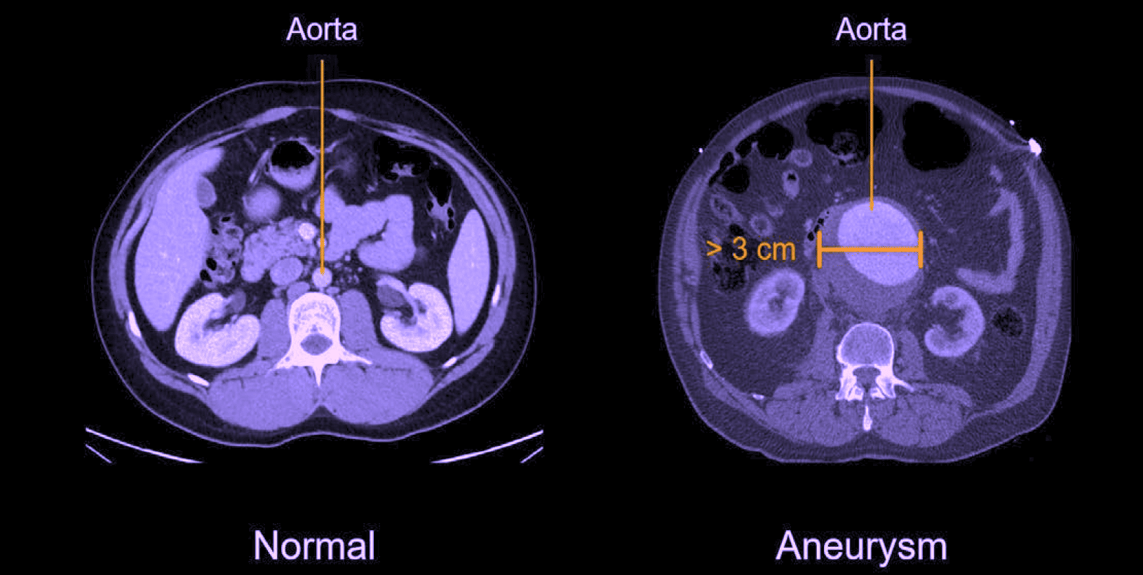
Computed tomography cross-sectional images of the abdominal aorta. On the left is a normal aorta with normal diameter. On the right is a dilated aorta (>3 cm), consistent with aortic aneurysm. The condition carries an increased risk of rupture and requires regular follow-up or treatment depending on size and growth.
When is CT of the aorta recommended?
A CT examination of the aorta is recommended when acute or chronic vascular changes are suspected. It is used both for diagnostics and to follow known conditions over time, especially in aneurysms where accurate size assessment is crucial.
Common symptoms and situations that may justify the examination:
- Sudden or persistent chest, back or abdominal pain.
- Suspected aortic aneurysm or dissection.
- Throbbing sensation in the abdomen.
- Follow-up of previously diagnosed aneurysm.
- Before or after surgical or endovascular treatment of the aorta.
- Investigation in cases of high cardiovascular risk, such as elevated blood lipids or smoking.
Conditions when CT Aorta is recommended
- Aortic aneurysm – dilation of the aorta that can pose a risk of rupture and requires regular follow-up.
- Aortic dissection – tear in the vessel wall that can be an acute life-threatening condition.
- Atherosclerosis – hardening of the arteries that can cause narrowing and affect blood flow.
- Intramural hematoma or bleeding in the aortic wall.
- Postoperative follow-up after vascular surgery or stenting.
- Traumatic vascular injuries after accidents.
- Congenital vascular anomalies that affect the structure and function of the aorta.
CT Aorta compared to MRI
CT of the aorta (CT angiography) has in many cases become the first-line method for investigating aortic diseases, especially in acute cases. Computed tomography is a rapid examination that provides very high resolution and makes it possible to accurately measure the diameter of the aorta and identify conditions such as aneurysm, dissection or bleeding.
Compared to MRI, CT provides better conditions for detecting acute changes and calcifications in the vessel wall. MRI can still be an alternative in some cases, for example when you want to avoid radiation, but CT is today the method most often used for both diagnosing and monitoring aortic aneurysms.
Order CT Aorta – we will send a referral directly
CT Aorta is a central examination for detecting and monitoring changes in the body's largest vessel. Using high-resolution images, the diameter of the aorta, the vessel wall and any abnormalities can be carefully analyzed, which is crucial for assessing risk and the need for treatment or follow-up.
The examination is performed using modern computed tomography and the results are always reviewed by a specialist in radiology. You will receive a written report that can form the basis for further medical assessment or treatment.
Note! The examination involves a low dose of ionizing radiation and is usually performed with contrast media for optimal image quality. Before the examination, consideration may need to be given to kidney function or any allergies to contrast media.


