
Quick version
What is a brain tumor?
A brain tumor is a broad collective name for several different tumor diseases that arise in the brain or nearby structures. A tumor is formed when a group of cells begins to divide in an abnormal and uncontrolled manner. Brain tumors can be both benign and malignant and differ greatly in terms of origin, growth rate, treatment and prognosis.
Primary brain tumors arise in the brain itself or its membranes. They should be distinguished from brain metastases, where a cancer that originated in another organ has spread to the brain. Examples of cancers that can relatively often cause brain metastases are lung cancer, breast cancer, kidney cancer and malignant melanoma.
Even a benign brain tumor can cause significant discomfort. The brain is located in a closed space inside the skull and a growing tumor can therefore press on or displace sensitive brain tissue even if the tumor cells do not grow invasively.
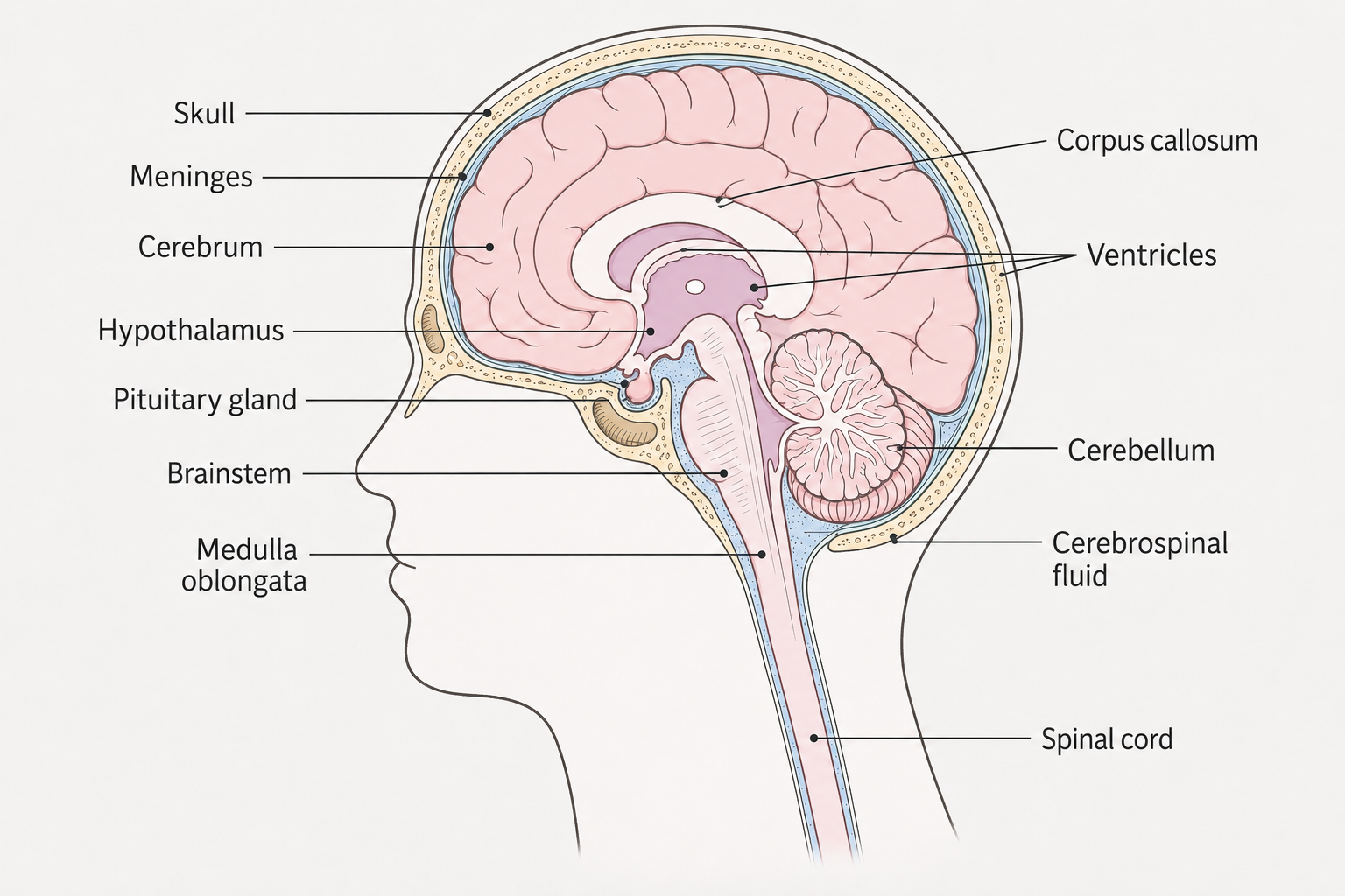
Illustration of the anatomy of the brain in cross-section, including the cerebrum, cerebellum, brainstem, pituitary gland, hypothalamus, ventricles and spinal cord. Brain tumors can arise in different parts of the brain and cause different symptoms depending on the location of the tumor and which structures are affected.
Different types of brain tumors
There are many different types of brain tumors. They are classified based on the cell type the tumor originates from, the tumor's molecular properties and how biologically aggressive it is.
Glioma
Glioma is a group of primary brain tumors that develop from glial cells or their precursors in the central nervous system. Glial cells normally have the task of supporting and protecting nerve cells and contributing to the normal function of the brain. Glioma is the most common form of malignant primary brain tumor in adults and includes several biologically different tumor types. The most important forms in adults are astrocytoma, oligodendroglioma and glioblastoma. Today, the classification is based not only on how the tumor cells look under a microscope but also on molecular and genetic properties. For example, changes in the genes and deletion of the chromosome arms are important for how different gliomas are classified, treated and assessed prognostically.
Gliomas are graded according to biological aggressiveness. Some low-grade gliomas can develop slowly over many years, while high-grade gliomas grow faster and infiltrate surrounding brain tissue. Glioblastoma is a grade 4 tumor and the most aggressive form of diffuse glioma in adults. Because gliomas often grow into the surrounding brain tissue, it can be difficult to remove all the tumor cells surgically.
Meningiomas
Meningiomas are tumors that arise from the meninges that surround the brain and spinal cord. They do not grow primarily from the brain tissue itself. Most meningiomas are slow-growing and are classified as WHO grade 1, but there are also more unusual grade 2 and grade 3 tumors with an increased risk of faster growth and recurrence. A meningioma may be asymptomatic and discovered by chance during MRI or CT scan performed for another reason. Symptoms occur primarily when the tumor becomes large enough to press on the brain, cranial nerves or blood vessels. Depending on the location, it can cause, for example, epileptic seizures, headaches, visual impairment, muscle weakness or other neurological symptoms.
Small meningiomas that do not cause symptoms do not always need to be treated but can be followed with recurrent imaging diagnostics. In the case of symptomatic or growing tumors, surgery or radiation therapy may be appropriate. Treatment is guided by, among other things, the size of the tumor, its location, growth and histological grade.
Medulloblastoma
Medulloblastoma is a malignant brain tumor that occurs primarily in children and usually arises in the cerebellum. The tumor can grow rapidly and has the ability to spread via the brain and spinal fluid to other parts of the central nervous system. In adults, medulloblastoma is considerably rarer. Treatment usually consists of surgery in combination with radiotherapy and chemotherapy. Diagnosis and treatment therefore differ clearly from the most common brain tumors in adults.
Other brain tumors
Other primary tumors of the central nervous system include ependymomas and germ cell tumors. Some tumor types occur primarily in children and younger people, while others are more common in adults and the elderly.
How common is a brain tumor?
Every year, about 1,300 people in Sweden are diagnosed with a brain tumor. The condition can occur at any age but is more common in older people and about half of those affected are over 60 years old. Brain tumors include several different tumor types with major biological differences. How the disease develops and is treated is based on the type of tumor. Statistics on survival and prognosis therefore always need to be interpreted based on the type of tumor.
Symptoms of a brain tumor
The symptoms of a brain tumor depend primarily on where in the brain the tumor is located, how quickly it grows, and whether it causes swelling or increased pressure in the skull. A slow-growing tumor may cause gradually increasing symptoms, while a more rapidly growing tumor may cause more pronounced symptoms for a shorter period of time. Symptoms and signs of a brain tumor may include:
- new epileptic seizures.
- headaches that are new or change over time.
- nausea and vomiting, especially with increased pressure in the skull.
- weakness or reduced mobility in an arm, leg, or one side of the body.
- numbness or altered sensation.
- balance problems or impaired coordination.
- vision changes or loss of visual field.
- speech difficulties or difficulties understanding language.
- memory problems or impaired cognitive ability.
- personality changes or changed behavior.
- pronounced fatigue or brain fatigue.
Headaches are common in the population and are very rarely due to a brain tumor. In the case of a brain tumor, headaches often occur together with other neurological symptoms or change in a clear way compared to the person's previous headaches.
Epileptic seizures can be a first symptom
A new epileptic seizure in an adult can be the first sign of a structural change in the brain and therefore needs to be investigated medically. A brain tumor can affect the brain's electrical activity and trigger both focal and generalized epileptic seizures. An epileptic seizure does not automatically mean that a brain tumor is present. There are many other causes of epilepsy, but new onset seizures are an important reason for neurological assessment and brain imaging.
Why does a brain tumor cause different symptoms?
Different parts of the brain control different functions. For example, a tumor in the frontal lobe can affect behavior, initiative, motor skills and/or language, while a tumor in the occipital lobe can affect vision. Changes near motor or sensory areas can cause weakness or loss of sensation, and tumors in the cerebellum can affect balance and coordination. Symptoms can also occur when the tumor causes swelling in the surrounding brain tissue or affects the circulation of cerebrospinal fluid. This can lead to increased pressure inside the skull.
What causes brain tumors?
In most cases, it is not possible to determine why an individual develops a primary brain tumor. The disease occurs through genetic and molecular changes in cells that alter the regulation of cell division and cell death. Most brain tumors are not directly hereditary. A small number of cases are linked to unusual genetic syndromes that increase the risk of tumors in the nervous system and other organs. Previous treatment with ionizing radiation to the head may also increase the risk of certain brain tumors later in life.
However, for the vast majority of people with brain tumors, there is no clear external cause that can be identified.
How is a suspected brain tumor investigated?
The investigation begins with a medical history and clinical examination. The doctor assesses, among other things, how the symptoms have developed and usually performs a neurological examination where, for example, reflexes, muscle strength, sensation, coordination, balance, vision and eye movements are checked.
If a change in the brain is suspected, imaging diagnostics are central. Computed tomography can quickly show larger tumors, bleeding, swelling, increased intracranial pressure and other acute changes. If a tumor is detected or the suspicion persists, the examination is usually supplemented with MRI for a more detailed assessment.
MRI brain for suspected brain tumor
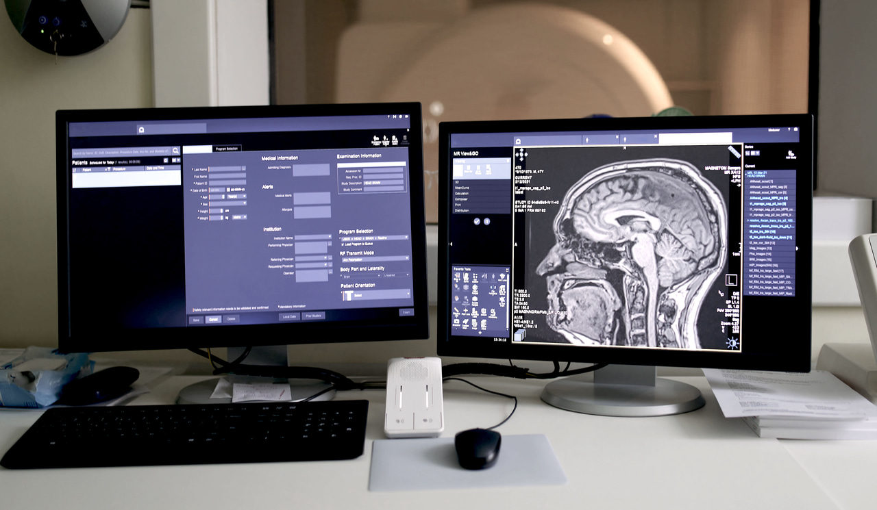
MR brain is the most important radiological method for detailed investigation and follow-up of brain tumors. MRI uses magnetic fields and radio waves and can image the soft tissues of the brain with high contrast without ionizing radiation. MRI can provide detailed information about the size of a change, its exact location, relationship to surrounding brain tissue and whether there is swelling around the tumor. The examination can also detect smaller or more difficult-to-identify changes that are not always visible as clearly with computed tomography.
In a specific tumor investigation, contrast agents are usually used to assess how the change absorbs contrast. Contrast enhancement and other MRI properties can provide important information about the nature of the tumor and help in planning for, for example, surgery. However, imaging cannot always determine with certainty the exact type of tumor.
In cases of strong clinical suspicion of a brain tumor, MRI may be warranted even if a previous computed tomography scan has not shown any clear tumor.
Computed tomography of the brain
Computed tomography, usually referred to as CT brain, is often used as the initial diagnostic imaging examination for acute or newly occurring neurological symptoms because the examination is quick and is available at most hospitals. CT can show, among other things, a larger tumor, bleeding, swelling, displacement of brain structures and signs of increased pressure in the skull. The method uses X-ray radiation and can be performed both with and without contrast agents, depending on the question.
If CT shows a possible brain tumor, the investigation is normally supplemented with MRI for more precise information about the extent and anatomical location of the tumor. MRI is therefore the first-line method for more detailed radiological investigation and follow-up of brain tumors.
MRI and CT complement each other
MRI and CT have different strengths and should not be seen as competing examinations. CT is particularly valuable when a rapid assessment is needed, for example in the case of acute neurological deterioration, suspected bleeding or severe pressure increase. MRI, on the other hand, provides more detailed information about brain tissue and the tumor's relationship to surrounding structures. Which examination is performed first therefore depends on symptoms, how urgent the situation is and the clinical question. In the case of a confirmed brain tumor that may be relevant for treatment, the investigation should usually include MRI unless there are medical contraindications to the examination.
Can MRI show if a brain tumor is cancer?
MRI can provide important information about how suspicious a change is by showing, among other things, the tumor's growth pattern, contrast uptake, surrounding swelling and relationship to brain structures. Some tumors have characteristic radiological features that allow a probable diagnosis to be made.
Important information about MRI and screening for suspected brain tumors
MRI brain is a very sensitive method for examining brain structures but is not used as a general screening for brain tumors in people without symptoms or medical issues. The examination can also detect changes that are benign or lack clinical significance and that may require further assessment.
If a brain tumor is suspected, the diagnosis is based on the combined assessment of symptoms, neurological examination, imaging diagnostics and, if necessary, tissue analysis. It is important to know that imaging cannot always reliably determine the exact type or biological properties of the tumor. For many brain tumors, analysis of tumor tissue is therefore needed to establish the diagnosis.
Biopsy and tissue analysis
A definitive tumor diagnosis is often based on histopathological and molecular analysis of tumor tissue. Tissue samples can be taken in connection with an operation where all or part of the tumor is removed or through a targeted biopsy if a larger operation is not appropriate. The pathologist analyzes the tumor cells and today often supplements the examination with molecular and genetic analyses. Such findings have become increasingly important for the classification of, for example, glioma and can affect both prognosis and choice of treatment.
Blood tests for brain tumors
There is no blood test or any general tumor marker that can diagnose or rule out a brain tumor. Blood tests are instead used primarily to assess general condition, organ function and possible alternative causes of symptoms, as well as to prepare for examinations and treatment. For certain specific and unusual tumor types, special laboratory tests or analyses of brain and spinal fluid may be relevant. Such sampling is done based on the specific clinical question.
Primary brain tumor and brain metastases
A primary brain tumor originates in the brain or its nearby tissues. A brain metastasis, on the other hand, occurs when cancer cells from a tumor elsewhere in the body spread to the brain.
The difference is important because treatment, prognosis and further investigation are guided by the original tumor. If brain metastases are suspected, the investigation may also include imaging of other parts of the body to identify or map the underlying cancer.
How are brain tumors treated?
Treatment depends on the tumor type, location, size, molecular characteristics, the patient's age and general condition. Treatment may consist of surgery, radiotherapy and drug therapy, alone or in combination.
Surgery
In many brain tumors, surgery is an important part of both diagnosis and treatment. The goal is usually to remove as much tumor tissue as possible without causing unacceptable damage to important neurological functions. The location of the tumor may therefore limit how much can be surgically removed.
Radiation therapy and drugs
Radiation therapy can be used after surgery, as the main treatment or in case of relapse depending on the type of tumor and the extent of the disease. Cytostatics and other drug treatments are used for certain types of tumors. Treatment is adapted to the biological and molecular properties of the tumor.
Cortisone can be used to reduce swelling around a tumor and antiepileptic drugs may be needed in people who have epileptic seizures.
Prognosis for brain tumors
The prognosis varies greatly between different types of brain tumors. A slowly growing and well-defined tumor can have a very good prognosis, while high-grade infiltrating tumors can be significantly more difficult to treat. The prognosis is affected by, among other things, tumor type, molecular properties, location, age, general condition and how much of the tumor can be removed or controlled with treatment. Overall survival statistics for all brain tumors are therefore difficult to use to assess the prognosis for an individual person.
When should you seek care?
Contact healthcare for new or gradually increasing neurological symptoms such as unexplained weakness, loss of sensation, balance difficulties, vision changes, speech difficulties, personality changes or a headache that has clearly changed character and persists.
A first epileptic seizure in an adult should always be assessed medically. Sudden onset of paralysis, difficulty speaking, impaired consciousness, severe acute headache or other rapidly developing neurological symptoms should be assessed urgently as they can also be caused by, for example, stroke or cerebral hemorrhage.