
Quick version
Myeloma is a chronic blood cancer that arises in the plasma cells in the bone marrow. The altered plasma cells can produce monoclonal immunoglobulins or free light chains and at the same time affect normal blood formation, the skeleton, the kidneys and the immune system. The investigation includes, among other things, M component, free immunoglobulin chains, blood status, calcium and kidney function tests together with bone marrow examination and imaging diagnostics. Treatment is adapted to the activity of the disease, genetic characteristics, kidney function, age and general condition.
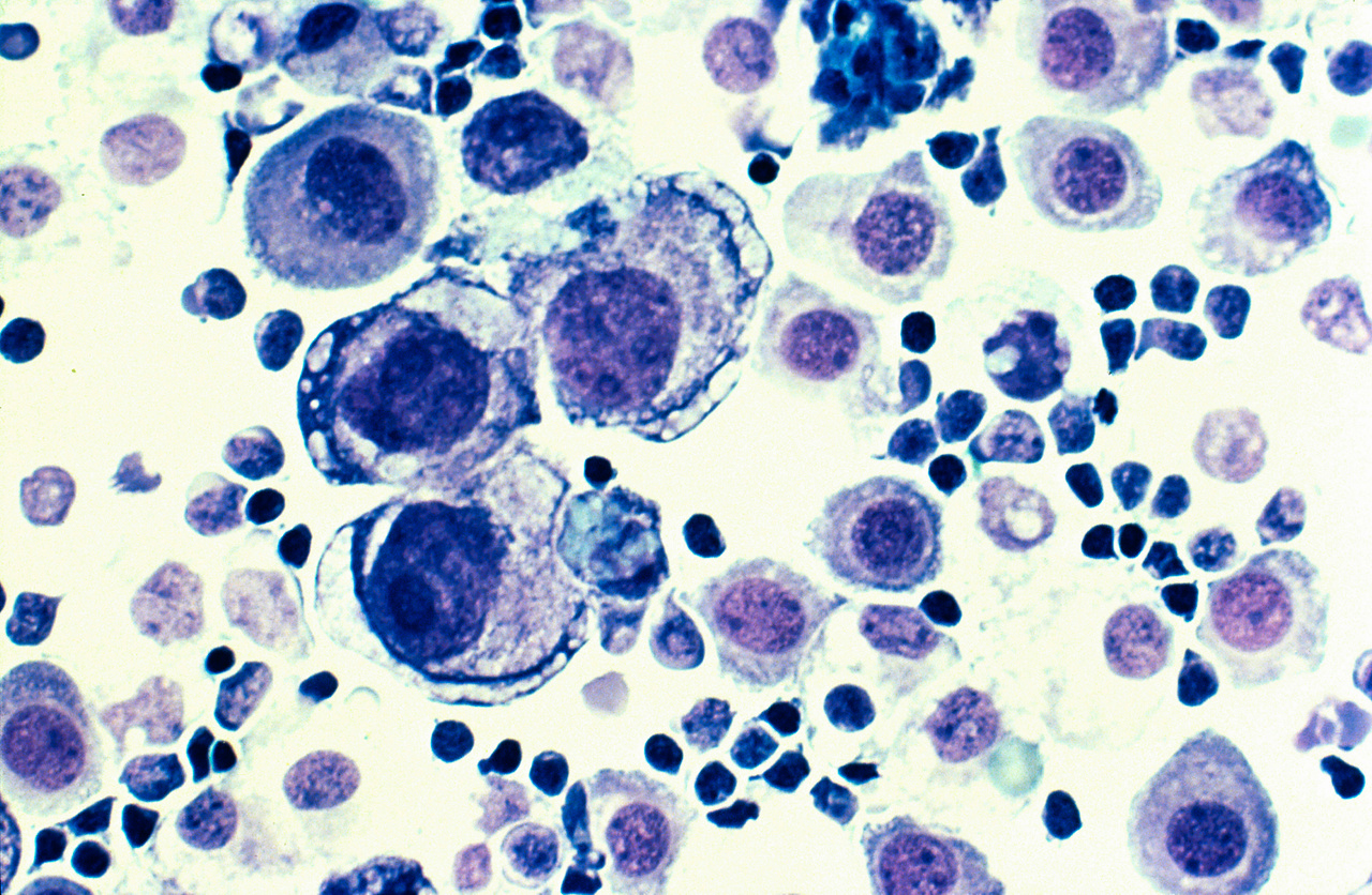
Myeloma, also known as multiple myeloma, is a form of blood cancer that occurs in the plasma cells in the bone marrow. Plasma cells are a type of white blood cell that normally produces antibodies, immunoglobulins, which help the body fight infections. In myeloma, a group of plasma cells changes and begins to divide uncontrollably.
The diseased plasma cells can crowd out the normal blood formation in the bone marrow and produce large amounts of the same antibody or parts of an antibody. This is called monoclonal production and can lead to anemia, impaired immune system, kidney damage and damage to the skeleton, among other things.
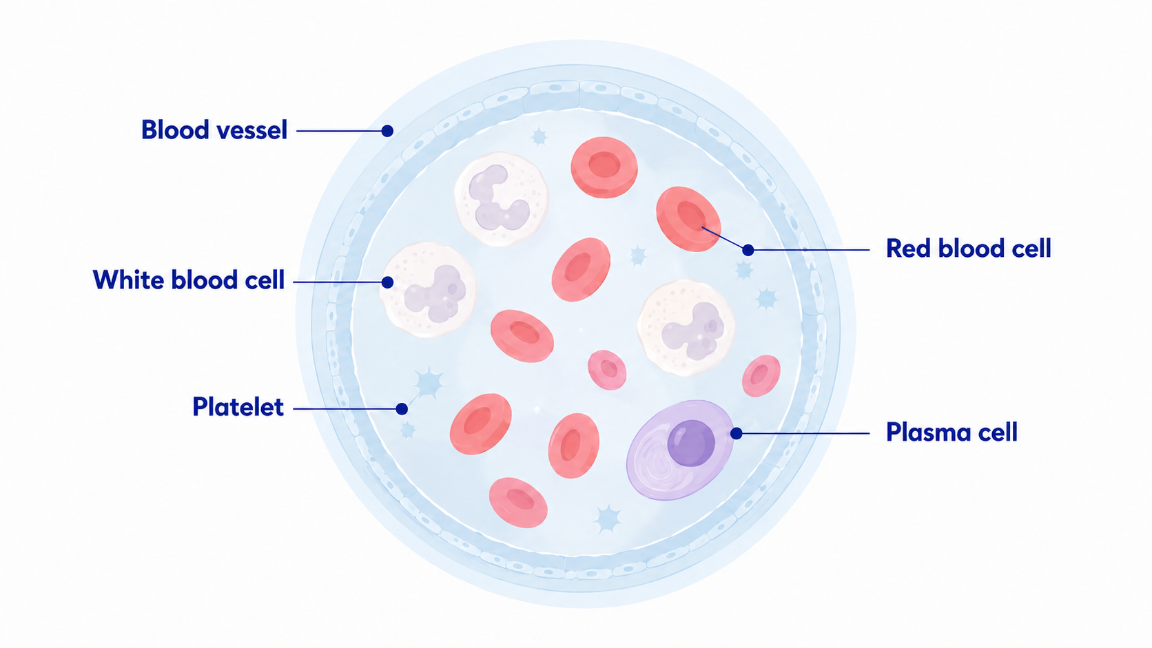
Illustration of blood cells with plasma cells, the type of cell that changes in myeloma.
Myeloma is a chronic cancer that often develops slowly. However, the course of the disease varies considerably between individuals and the need for treatment depends, among other things, on how much the disease affects the bones, kidneys, blood formation and other organs.
How common is myeloma?
In 2025, 894 people were diagnosed with myeloma in Sweden, of whom 512 were men and 382 were women. The disease occurs primarily in older people and is uncommon before the age of 40. Survival has improved significantly in recent decades in line with the development of new drugs and more effective combination treatments.
How does myeloma occur?
Myeloma occurs when a plasma cell in the bone marrow undergoes genetic changes that cause it to begin to divide uncontrollably. The new plasma cells are genetically related and form a clone of cells that produce the same type of immunoglobulin or the same type of free immunoglobulin light chain.
The monoclonal production can often be measured in blood or urine as a so-called M component. In some people, myeloma cells produce mainly free light chains of the kappa or lambda type, which can be detected by analysis of Free immunoglobulin chains (FLC).
Symptoms of myeloma
Myeloma often takes a long time to develop and may cause few or no symptoms in the early stages. The disease is therefore sometimes discovered in connection with blood tests or other examinations carried out for another reason. When symptoms occur, they are often due to the effects on the skeleton, blood formation, kidneys or immune system.
Common symptoms and findings of myeloma may include:
- bone pain, especially in the back, ribs or pelvis.
- increased risk of fractures as the bones weaken.
- fatigue and decreased energy due to anemia.
- recurrent or unusually severe infections.
- decreased kidney function.
- foamy urine or swelling if the kidneys are affected.
- increased thirst, nausea or confusion if calcium levels are elevated.
- numbness, loss of sensation or muscle weakness if the nerves or spinal cord are affected.
- unintentional weight loss and worsening general condition.
The symptoms are not specific to myeloma and can have many other causes. The combination of, for example, unexplained anemia, bone pain, kidney involvement, elevated calcium or an M component may, however, justify further medical investigation.
Why are the bones affected in myeloma?
The myeloma cells affect the balance between the cells that build and break down bone tissue. The breakdown of bone increases while new formation does not compensate for the loss. This can lead to areas of weakened bone tissue, so-called osteolytic changes.
Skeletal involvement can cause long-term pain, vertebral compressions and an increased risk of fractures. The back and chest are common areas of discomfort. If changes in the spine affect nerve roots or the spinal cord, numbness, muscle weakness and other neurological symptoms can also occur.
Myeloma and M component
Myeloma cells often produce large amounts of an identical immunoglobulin. This monoclonal protein is called the M component and can be identified by protein analyses in blood and sometimes urine.
Serum protein fractions or serum electrophoresis are used to identify and measure any M component. Immunofixation can then be used to determine the type of immunoglobulin and light chain produced.
An M component does not automatically mean that a person has myeloma. It can also occur in, for example, MGUS and other monoclonal gammopathies. The result therefore always needs to be assessed together with other laboratory tests, bone marrow examination and possible organ involvement.
Free immunoglobulin chains in myeloma
Free immunoglobulin chains are an important analysis in the investigation and follow-up of myeloma and other monoclonal plasma cell diseases. The analysis measures free immunoglobulin light chains of the kappa and lambda types and the ratio between them.
In a person without monoclonal plasma cell disease, both kappa and lambda are produced in relative balance. If a clone of plasma cells begins to produce large amounts of only one type of light chain, the level of kappa or lambda can rise sharply and the kappa/lambda ratio becomes clearly abnormal.
S FLC Kappa shows the concentration of free kappa chains while S FLC Lambda shows the concentration of free lambda chains. shows the ratio between the two chains and can provide important information about possible monoclonal production.
However, an abnormal ratio alone cannot establish the diagnosis of myeloma. For example, impaired kidney function can increase both kappa and lambda and therefore needs to be taken into account when interpreting.
What is light chain myeloma?
In light chain myeloma, the plasma cells mainly produce free light immunoglobulin chains instead of a complete immunoglobulin. The chains can be of the kappa or lambda type and are present in both blood and urine.
In this form of myeloma, a traditional serum electrophoresis may show little or no clear M component, while analysis of free light chains shows a sharp increase in the involved chain and a clearly abnormal kappa/lambda ratio.
What blood tests are used when myeloma is suspected?
The investigation of suspected myeloma includes several laboratory tests. The purpose is both to identify monoclonal plasma cell production and to assess whether the disease has affected blood formation, the kidneys, the skeleton or other organs.
Relevant analyses may include:
- Free immunoglobulin chains (FLC) for analysis of kappa, lambda and the kappa/lambda ratio.
- serum protein fractions or serum electrophoresis to identify and quantify the M component.
- immunofixation to determine the type of monoclonal immunoglobulin present.
- immunoglobulins IgG, IgA and IgM for assessment of antibody production.
- Hemoglobin and other blood status to identify, for example, anemia.
- Calcium because bone breakdown can cause hypercalcemia.
- Creatinine and eGFR to assess kidney function.
- Albumin which is used, among other things, in assessment and staging.
- Beta 2 microglobulin which is an important prognostic marker in myeloma.
- Lactate dehydrogenase which can provide additional prognostic information.
RCC recommends, among other things, blood count, calcium, creatinine, albumin, fractionated proteins and free light chains when myeloma is suspected. Which tests are needed is determined by the clinical question.
Urine test in myeloma
Urine tests can be an important part of the investigation because free immunoglobulin light chains can be filtered through the kidneys and appear in the urine. Historically, these chains are sometimes called Bence Jones protein.
Urine analysis can be used to identify and classify monoclonal proteins. Free light chains in serum can supplement or replace certain urine analyses in some parts of the investigation, but when myeloma is confirmed, urine samples can still play an important role in diagnosis and follow-up.
Calcium in myeloma
Calcium is an important blood test in suspected or confirmed myeloma. When the disease causes increased bone breakdown, calcium can be released from the skeleton and the concentration in the blood rises. This is called hypercalcemia.
Elevated calcium can cause thirst, increased urine production, constipation, fatigue, muscle weakness and, at more pronounced levels, confusion or impaired consciousness.
Renal effects in myeloma
The kidneys can be affected in several ways in myeloma. Free light chains can be filtered through the kidneys and in high concentrations cause damage to the renal tubules. Hypercalcemia, dehydration, infections and certain medications can also contribute to impaired renal function.
Creatinine and eGFR are therefore used to assess renal function. Renal values are also important when interpreting free light chains because both kappa and lambda can increase when the kidneys' filtration capacity deteriorates.
Kidney involvement can sometimes cause symptoms such as foamy urine, swelling and shortness of breath, but impaired kidney function can also occur without clear symptoms.
Anemia and blood status in myeloma
Myeloma cells can take up space in the bone marrow and inhibit normal blood formation. A common consequence is anemia, i.e. low hemoglobin, which can cause fatigue, weakness, shortness of breath and reduced physical performance.
In more extensive bone marrow involvement, the number of white blood cells and platelets can also be affected. A lack of normal immune cells can increase the risk of infection, while low levels of platelets can increase the tendency to bleed.
How is myeloma diagnosed?
The investigation of suspected myeloma is based on a combination of blood tests, urine tests, bone marrow examination and diagnostic imaging. No single examination can establish the diagnosis alone.
Blood tests are used, among other things, to identify M component and free light chains and to assess blood status, calcium and kidney function. Urine tests can provide additional information about monoclonal proteins and excretion of free light chains.
Bone marrow tests are used to assess the amount of clonal plasma cells and their biological and genetic properties. Imaging diagnostics are also used to map the impact on the skeleton and bone marrow. The final diagnosis is made within hematological specialist care.
CRAB criteria for myeloma
In diagnostics, the term CRAB is often used to describe classic forms of organ involvement that can be caused by myeloma.
- C stands for calcium and refers to elevated calcium in the blood.
- R stands for renal impairment and refers to impaired kidney function.
- A stands for anemia and refers to anemia.
- B stands for bone lesions and refers to myeloma-related skeletal changes.
The presence of one or more of these findings, together with signs of clonal plasma cell disease, may indicate myeloma requiring treatment. At the same time, it is important that organ involvement is actually assessed to be due to the myeloma disease and not to any other cause.
Myeloma can be diagnosed before clear symptoms appear
Myeloma does not always have to have caused clear organ involvement for treatment to be relevant. The diagnostic criteria also include certain laboratory and imaging findings that imply a very high risk of organ damage developing. For example, this may involve a very high proportion of clonal plasma cells in the bone marrow, severely abnormal free light chains under certain conditions or specific findings on MRI. These criteria are assessed within specialist care and cannot be interpreted based on a single blood sample.
Bone marrow test in myeloma
Bone marrow examination is a central part of the diagnosis. A sample is usually taken from the pelvic bone and analysed to determine the proportion of bone marrow cells that are clonal plasma cells.
The myeloma cells can also be analysed genetically, for example with FISH, to identify chromosomal changes that can affect risk classification, prognosis and treatment strategy.
Computed tomography and MRI in myeloma
Imaging is an important part of the investigation of suspected myeloma because the disease can cause changes in both the skeleton and the bone marrow. In Swedish myeloma investigations, low-dose computed tomography of the myeloma skeleton is the standard method for mapping osteolytic changes and other skeletal lesions. Computed tomography has high sensitivity for destruction in the skeleton and can also show plasmacytomas outside the bone marrow.
MRI is not routinely used as a general whole-body examination in suspected myeloma. However, the method is particularly sensitive for changes in the bone marrow and is used for specific clinical questions. MRI is the first-line examination in suspected involvement or compression of the spinal cord and can also be used in the investigation of solitary skeletal plasmacytoma.
In asymptomatic myeloma, MRI of the spine can be used as a supplementary examination to identify focal changes in the bone marrow that may affect the assessment of whether the disease requires treatment. Which imaging method is used is therefore determined by the clinical situation and the results of other myeloma investigations.
The imaging is assessed together with blood tests, urine tests and bone marrow examination. No single imaging examination can alone confirm or exclude myeloma.
MGUS and smoldering myeloma
Myeloma is often preceded by an earlier stage of monoclonal plasma cell production. In MGUS, monoclonal gammopathy of undetermined significance, there is a small amount of monoclonal protein or clonal plasma cells without the criteria for myeloma being met.
MGUS becomes more common with increasing age and is often discovered incidentally in connection with blood tests. Most people with MGUS never develop myeloma, but the condition is followed because the risk of progression is higher than in people without monoclonal gammopathy.
Smoldering myeloma, also called asymptomatic myeloma, is between MGUS and myeloma requiring treatment. The amount of monoclonal protein or clonal plasma cells is greater than in MGUS, but the disease has not yet caused the findings that require treatment. The risk of progression is therefore higher and regular follow-up is important.
How is myeloma treated?
Treatment is tailored to the activity of the disease, genetic characteristics, the patient's age, general condition, kidney function and other medical factors. In asymptomatic myeloma, regular follow-up may be sufficient, while myeloma requiring treatment is usually treated with a combination of several drugs.
Treatment may include proteasome inhibitors, immunomodulatory drugs, monoclonal antibodies, cortisone and other targeted treatments. For patients who are deemed suitable, high-dose treatment followed by autologous stem cell transplantation may be included.
In the event of relapse, there are additional treatment options and for some patients, newer immunotherapies such as bispecific antibodies or CAR T cell therapy may be considered. The choice of treatment is made within specialist hematological care.
Treatment of skeletal involvement and other complications
In addition to treatment for the myeloma disease itself, treatment is often needed to prevent or treat complications. Drugs that reduce bone breakdown can be used to reduce the risk of fractures and bone pain.
Pain relief, treatment of infections, treatment of hypercalcemia and support for impaired kidney function can also be important parts of care. In the case of localized painful bone changes or plasmacytoma, radiation therapy may be appropriate.
Prognosis for myeloma
The prognosis for myeloma varies considerably between individuals and is influenced by factors such as age, general condition, kidney function, the genetic characteristics of the disease, stage and how well the disease responds to treatment.
Treatment options have improved significantly and many people can live with the disease for a long time with recurring treatment periods. The relative ten-year survival rate in Sweden is around 37 percent, but population statistics cannot be used to predict the prognosis for an individual person.
When should you seek medical attention?
Contact your healthcare provider if you have persistent and unexplained bone pain, especially in the back or chest, severe fatigue, recurrent infections, unexplained anemia, worsening kidney function or other new symptoms that do not go away.
Sudden severe back pain along with muscle weakness, numbness, loss of sensation, or trouble controlling urine or stool should be evaluated urgently as these may be signs of spinal cord involvement.
Important about blood tests and screening
Free light chains, M component, calcium, and other blood tests can provide important information when myeloma is suspected but should not be used as stand-alone screening tests for cancer in people without medical concerns. An abnormal test result does not automatically mean myeloma, and normal values alone cannot rule out the disease.
If myeloma is suspected, the combined assessment of symptoms, laboratory tests, urine tests, bone marrow examination, and imaging tests determines whether further investigation is needed.