


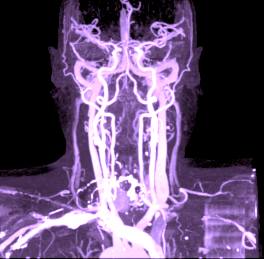
Computed tomography of the blood vessels of the neck
CT carotid 3D angio is an X-ray examination used to visualize the blood vessels in the neck, especially the carotid arteries (carotid arteries) and vertebral arteries that supply the brain with blood. The examination is performed using computed tomography and contrast media and is also called CT angiography (CTA) of the carotid arteries.
During the examination, an intravenous contrast agent is given that makes the blood vessels appear clearly on the X-ray images. Using advanced image reconstruction, the vessels can then be visualized in three dimensions (3D angiography). This makes it possible to accurately assess the anatomy of the vessels, detect narrowing, plaque formation or other vascular changes that can affect blood flow to the brain.
What does 3D angiography mean?
3D angiography means that the cross-sectional images of the computed tomography are reconstructed into three-dimensional models of the blood vessels. This allows the doctor to analyze the structure of the vessels from several different angles and get a more detailed picture of any changes.
The three-dimensional visualization makes it possible to:
- Assess the anatomy and course of the vessels more precisely.
- Identify narrowings (stenoses) in the carotid arteries.
- Detect atherosclerotic plaques and vascular changes.
- Assess the degree of any vascular narrowing.
- Identify vascular abnormalities such as dissection or aneurysm.
This type of advanced image analysis makes CT carotid angiography one of the most reliable non-invasive methods for mapping the blood vessels of the neck.
What can the examination show?
CT angiography of the carotid arteries is primarily used to detect changes in the arteries that transport blood to the brain. The examination may show, among other things:
- Narrowings in the carotid arteries (carotid stenosis).
- Atherosclerotic plaques in the carotid arteries.
- Vascular dissection (damage to the vessel wall).
- Aneurysm or other vascular abnormalities.
- Changes in the vertebral arteries.
Narrowings in the carotid arteries are often caused by hardening of the arteries (atherosclerosis). If a narrowing becomes pronounced, blood flow to the brain can be affected and the risk of transient ischemic attack (TIA) or stroke can increase.
By visualizing both the lumen of the vessels and any plaque formation, CT carotid angiography can provide a detailed picture of the condition of the vessels and help identify people at increased risk of cerebrovascular events.
When can a CT carotid angiography be medically justified?
The examination is often used when there is suspicion of disease in the blood vessels of the neck or when a more detailed mapping of the vessels is needed. It may be relevant, for example, in the following cases:
- Suspected narrowing of the carotid arteries.
- Investigation after TIA or stroke.
- Neurological symptoms such as sudden dizziness, visual disturbances or transient weakness.
- Suspected vascular dissection in the carotid arteries.
- Follow-up of previously detected vascular changes.
The examination can also be used to map the vascular anatomy prior to surgical or endovascular treatments.
After the examination, the images are reviewed by a specialist in radiology. The results are compiled in a radiological report in which any vascular changes, plaques or narrowing are described and graded.


