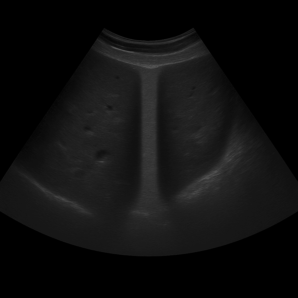
A biliary tract ultrasound is primarily performed to examine the bile ducts and their connection to the liver and gallbladder. The examination is performed by a radiology specialist and provides detailed real-time images that show whether there are obstructions, dilations or other changes in the biliary tract system. A biliary tract ultrasound is often used when there is suspicion of gallstone disease, blockage of bile flow or an impact on the outflow of the liver.
Biliary tract ultrasound – for abdominal pain, jaundice or elevated liver values
A biliary tract ultrasound is recommended for pain under the right rib cage, jaundice (yellow skin or whites of the eyes), nausea or itching of the skin. The examination is also used when blood tests show elevated liver or bile values, which may indicate obstruction of bile flow or inflammation. It is also valuable in the follow-up of previous biliary tract disease or surgical procedures.
Unlike MRI and CT, which are used for more detailed mapping of the course of the bile ducts or suspected tumors, ultrasound is the first-line method for suspected mechanical obstruction, gallstones or inflammation. The method is fast, radiation-free and shows changes in real time – for example, how the bile ducts widen when blocked or how bile flow is affected.
Common symptoms and questions
- Pain or pressure under the right rib cage.
- Jaundice or itching (signs of impaired bile flow).
- Elevated liver or bile values in blood tests.
- Suspected gallstones stuck in the bile duct.
- Follow-up after biliary surgery or previous biliary tract disease.
- Suspected inflammation, cysts or tumor changes in the bile ducts.
Conditions that can be detected with biliary tract ultrasound
- Bile duct dilation (dilation) due to obstruction or stone.
- Gallstones (choledochal calculi) in the major bile ducts.
- Inflammation of the bile ducts (cholangitis).
- Narrowing or blockage of bile flow.
- Cysts, tumors or other changes in the bile ducts.
- Consequences of surgery or infection of the bile ducts.
How an ultrasound of the biliary tract is performed
The examination is performed while you lie on your back or slightly on your left side. A gel is applied to the skin and the doctor moves the ultrasound probe over the upper part of the abdomen, just below the rib cage. The examination usually takes 15–20 minutes and requires you to fast for a few hours beforehand so that the bile ducts can be clearly assessed.
Order an ultrasound examination of the bile ducts – get a report and recommendation from a doctor
The images are reviewed by a specialist in radiology who draws up a written medical report. The answer is delivered digitally within a few working days and can be shared with your treating doctor for further investigation or treatment. If necessary, the examination can be supplemented with MRI or CT for a more detailed mapping of the bile duct system.

