
Quick version
To many, CT and MRI sound like two versions of the same thing: a machine that takes pictures of the body. But when a doctor chooses between them, it's not just about image quality. It's about what question needs to be answered, how quickly the answer is needed, whether the examination involves radiation, and whether the patient, for example, has metal implants, kidney disease, or severe claustrophobia.
For someone who has received a referral, it can therefore feel confusing when someone says that CT is best in one situation and MRI in another. The difference between the methods becomes clearer when you see what they actually show – and why the right method is often part of the diagnosis itself.
What are CT and MRI, and what are the differences between the technologies?
CT stands for computed tomography. The method uses X-rays to create cross-sectional images of the body. The images are taken quickly and are very good for showing bones, bleeding, lungs, and many acute conditions. In practice, CT is often the first choice when time is of the essence, for example in the case of a suspected stroke, head injury or internal bleeding.
MRI stands for magnetic resonance imaging, or magnetic resonance tomography. It uses strong magnetic fields and radio waves, not ionizing radiation. MRI provides particularly detailed images of soft tissues such as the brain, spinal cord, muscles, ligaments, joints and certain organs. Therefore, MRI is often used when you need to see tissues that do not appear as clearly on CT.
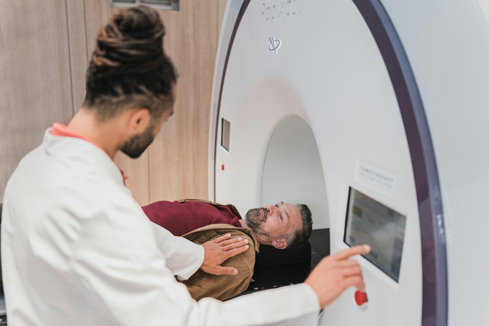
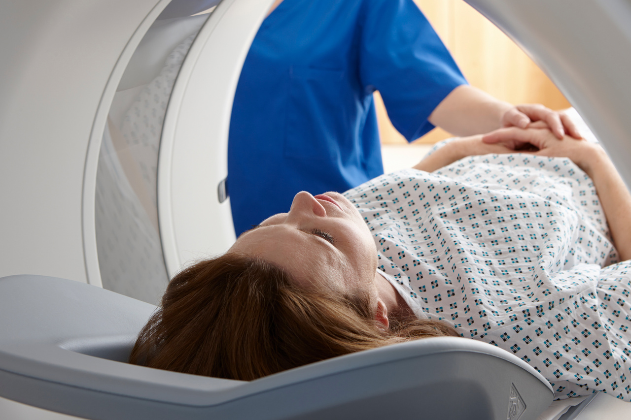
The difference in technique also affects the feel of the examination. A CT usually takes a few minutes, while an MRI usually takes longer and requires you to lie still in a narrower tunnel. For some patients, it is a small detail, for others it is absolutely crucial for which examination works in practice.
When do you choose CT over MRI?
CT is often chosen when the answer is needed quickly. In acute head injuries, CT is the first choice method because it can quickly show fresh bleeding and skull fractures. In suspected stroke, computed tomography is used early to distinguish between cerebral hemorrhage and blood clot, as this difference directly guides treatment.
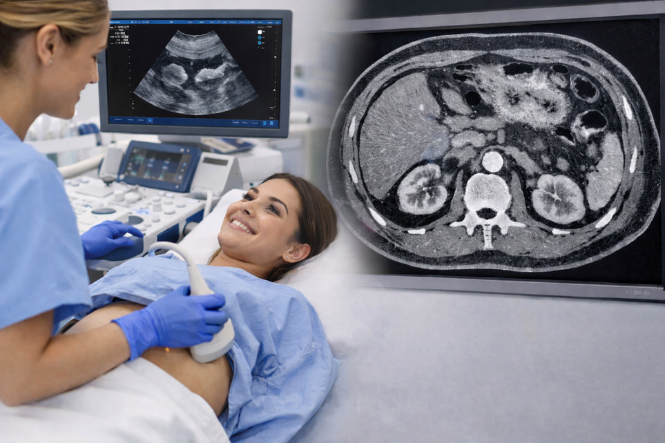
CT is also very useful in acute abdominal complaints. Examples include suspected appendicitis with complications, intestinal obstruction, kidney stones or suspected internal bleeding after trauma. The method provides a quick overview of several organs at the same time, which is a great advantage in the emergency department.
Lungor is another area where CT is often superior. In lunginflammation with complications, pulmonary embolism or investigation of pulmonary nodules, CT provides detailed images that MRI can rarely replace. Skeletal injuries, especially complex fractures, are also often best assessed with CT.
In some situations, the question is not “which examination is the most advanced?” but “which examination gives the right answer the fastest?”. In this case, CT often wins.
When is MRI the better choice?
MRI is often best when more information about soft tissues is needed. In cases of long-term back pain with suspected herniated disc affecting nerve roots, inflammation of the spine or changes in the spinal cord, MRI is often more accurate than CT. The same applies to many knee, shoulder and hip problems where one wants to assess meniscus, cruciate ligament, cartilage or muscle tendons.
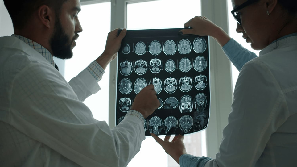
The brain is a classic example. MRI can show small changes that are not always visible on CT, for example some tumors, inflammations, multiple sclerosis and smaller areas of oxygen deficiency. Therefore, MRI is often used when the patient has neurological symptoms that are not explained by a quick emergency CT.
MR is also important in cancer investigation and follow-up in selected organs. Liver, pelvis, prostate, uterus and certain soft tissue tumors can sometimes be assessed in more detail with MRI. The examination then provides better contrast between different tissues and can help the doctor determine whether a change appears benign or more suspicious.
At the same time, this does not mean that MRI is always “better”. A method may be more detailed but still less suitable if it takes too long, is difficult to perform or does not best answer the clinical question.
Radiation, contrast agents and safety – what do you need to know?
The clearest medical difference is that CT uses ionizing radiation, while MRI does not. The radiation dose from a single CT is usually medically justified and the benefit then outweighs the risk, but unnecessary CT examinations are avoided – especially in children, younger people and in conditions where repeated examinations may be needed.
Both CT and MRI can be performed with contrast agents to make certain structures clearer. CT usually uses iodine-based contrast agents. It may be unsuitable for certain degrees of kidney damage and can in rare cases cause allergic or allergy-like reactions. MRI usually uses gadolinium-based contrast agents, which generally cause fewer allergic reactions but require special caution in severe kidney disease.
MRI instead has its own safety issues. The magnetic field means that certain implants, metal fragments or older types of pacemaker equipment may be an obstacle or require special checks before the examination. Many modern implants are MRI-conditional, but this must always be verified before booking.
For pregnant women, people with impaired kidney function or patients who have previously reacted to contrast, an individual assessment is made. That is why you often have to answer questions about kidneys, allergies, surgeries and metal in the body before the examination.
How do you know which method to choose?
The short answer is that the patient should rarely have to choose between CT and MRI. It is the medical question that determines. The doctor weighs together symptoms, status, blood tests, age, time course, previous illnesses and what you actually need to see in the images.
A few typical examples make the choice more understandable:
Sudden hemilateral weakness and speech difficulties: often acute CT first, sometimes followed by MRI.
Knee pain after twisting without a clear fracture on X-ray: often MRI if meniscus or ligament damage is suspected.
Suspected kidney stone with severe flank pain: often CT.
Long-lasting headache with neurological symptoms without acute trauma: often MRI.
Suspected internal bleeding after an accident: often CT.
Suspected nerve damage from a herniated disc: often MRI.
Another important question is whether imaging is needed at all. For example, not all back pain should be investigated with MRI, and not all headaches require CT. Radiology guidelines emphasize that the correct examination may also be to forgo imaging diagnostics when symptoms and examination findings do not indicate a serious illness. This reduces the risk of unnecessary findings that create concern without improving care.