
Quick version
Concussions are rarely seen on standard MRI because they are primarily functional disorders. MRI is therefore used in boxing to detect other silent, structural residual injuries — such as microbleeds and tissue changes — and to create a comparable baseline. In acute symptoms after a concussion, CT (computed tomography) is the first choice, while MRI is a crucial support in long-term safety work to assess the risk of continued exposure.
A hard hit to the chin can cause more than a few seconds of instability. For a boxer, repeated blows to the head can leave marks that are not visible from the outside, even when the person feels ready to continue training. Therefore, the question of a brain MRI often arises in connection with licensing, medical check-ups or after a knockout.
Why do boxers need to undergo a brain MRI?
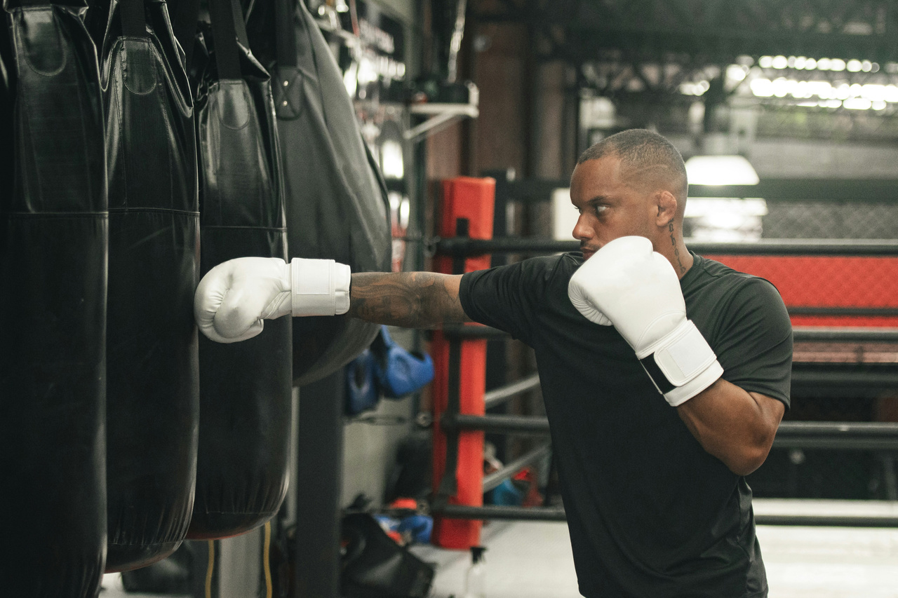
Boxing is different from many other sports because the head is a direct target for blows. This makes the risk of both acute and long-term brain damage medically relevant, even in well-trained people without obvious symptoms. Several boxing rules and medical minimum requirements use brain MRI as part of safety work, including as a baseline examination or recurring check-ups for competing boxers.
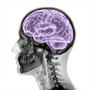
MRI, or magnetic resonance imaging, uses strong magnetic fields and radio waves to create detailed images of the brain without ionizing radiation. The examination can show structural changes that a standard neurological examination does not always pick up, such as old bleeding, scar-like changes or signs of tissue damage after previous trauma.
MRI brain is used to find injuries that are not immediately noticeable
A boxer can feel “clear-headed” and still carry changes after previous blows. MRI brain is therefore used not only when someone is feeling acutely unwell, but also to assess whether there are silent structural injuries that may increase the risk of continuing combat sports. This is particularly relevant after knockouts, repeated matches in a short period of time or recurrent head trauma during sparring and competition.
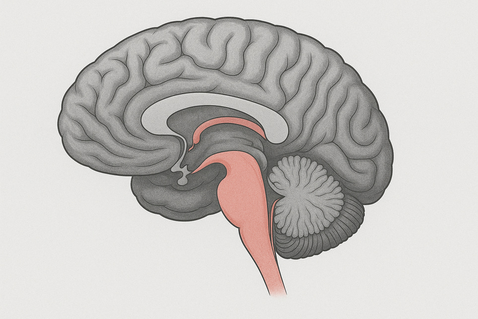
On MRI, the doctor can sometimes see things like small microbleeds, contusion residues or changes in the brain's white matter. Some advanced MRI sequences are more sensitive than CT scans for subtle residual injuries after trauma, especially in the subacute or chronic stage when symptoms are no longer dramatic but question marks remain.
This does not mean that MRI always shows an injury after a blow to the head. In sports-related concussions, standard MRI is often normal, since concussions are primarily a functional disorder rather than a clear structural injury on standard images. This is precisely why MRI is most valuable when you want to rule out more serious changes or assess consequences after repeated exposure.
Concussions are not the same as bleeding or permanent brain damage
Many people mistakenly believe that a concussion is always visible on MRI, but this is usually not the case. A concussion can cause headaches, nausea, dizziness, sensitivity to light and sound, slower thinking or memory impairment, but standardized imaging often shows normal results despite clear symptoms.
The medical purpose of MRI brain in boxers is therefore broader than "proving a concussion". The examination is used to look for things that change the risk picture: previous bleeding, scar tissue, deformities, atrophy or other findings that can make continued boxing more dangerous. For example, if a boxer has persistent neurological symptoms weeks after a match, or develops new symptoms after a relatively light hit, the need for in-depth investigation becomes greater.
In the acute stage, however, computed tomography, CT, is often used first if fresh bleeding or a serious head injury is suspected. CT is usually the first choice for acute mild head trauma when imaging is needed, while MRI has a greater role in persistent or unexplained symptoms later in the course.
Repeated blows to the head can cause cumulative risk
What makes boxing special is not just the individual knockout, but the cumulative exposure. Research on boxers and other martial artists shows that repetitive head blows can be linked to changes in brain structure and function over time, even when each individual occasion did not lead to a diagnosed concussion.
Studies of professional fighters have shown a connection between longer exposure, more fights or more knockouts and reduced volume in certain brain regions as well as changes in white matter. This does not mean that all boxers develop permanent brain damage, but it strengthens the case for regular medical monitoring in a sport where stress is directed at the brain.
There is also great interest in chronic traumatic encephalopathy (CTE), a neurodegenerative disease linked to repeated head trauma. CTE cannot currently be diagnosed with a standard MRI, but can only be reliably determined by examining brain tissue after death. However, MRI can show other structural damage after trauma, which gives the doctor an important basis for assessing whether it is safe to return to activities with a risk of new head impacts.
Therefore, MRI may be required even without clear symptoms
A common objection is that a healthy boxer without any problems should not need the examination. However, in boxing, medical rules are not only about current health, but about reducing the risk of catastrophic injuries in the next match. Therefore, there are requirements for baseline MRI or annual MRI checks in some professional regulations and for some ranked boxers.
A baseline examination makes it easier to compare future images. If a boxer later develops symptoms, is knocked out or needs a new license assessment, the doctor can compare with previous MRIs and see if anything has changed. It is especially valuable when you want to distinguish old, already known findings from new changes.
MRI can also detect conditions that are not caused by boxing but still affect safety, such as vascular malformations or other structural abnormalities. Such a finding does not necessarily mean illness in everyday life, but it can be crucial when the person is exposed to repeated blows to the head.
When should a boxer seek medical attention quickly after a blow to the head?
Not all symptoms after a match are “normal boxing symptoms”. Some signs should be taken seriously immediately, as they may indicate bleeding or other serious brain damage. Examples include:
- increasing headaches
- repeated vomiting
- confusion or difficulty waking up
- worsening memory lapses
- balance problems or slurred speech
- weakness, numbness or cramps
- unconsciousness, even for a short time
Such symptoms require urgent medical assessment. In such cases, the goal is first to detect or rule out a recent serious injury, often with CT in emergency care, and then to determine whether further investigation with MRI or neurological follow-up is needed.
If the symptoms have instead become prolonged, for example persistent headaches, difficulty concentrating, poor reaction time, unusual fatigue or personality changes weeks after a blow to the head, it is also wise to seek medical attention. The question then often becomes not just whether it is a transient concussion, but whether there is reason to pause boxing and do a more complete neurological assessment.
MRI brain scans in boxers are therefore less about routine for the sake of routine and more about making safer decisions when much is at stake. A normal examination does not give a free pass for unlimited exposure, but it can be an important support when doctors, trainers and boxers need to weigh performance against long-term brain health.