Quick version
Computed tomography (CT) creates detailed cross-sectional images of the body to quickly detect diseases and injuries. The examination is painless and often takes a few minutes. Sometimes contrast agents are given for clearer images, which requires extra monitoring of kidney function and a temporary interruption of the diabetes medication metformin. The radiation dose is higher than with a regular X-ray, but the examination is only done when the medical benefit outweighs the risk.
A CT scan, or computed tomography, is often ordered when the answer needs to come quickly and the images need to show more than a regular X-ray. This could be a sudden headache where bleeding needs to be ruled out, a suspected pulmonary embolism, kidney stones with severe pain, or an abdomen where the doctor wants to see if there is inflammation, blockage, or bleeding. For many, the examination itself is unknown territory, and then the questions tend to be less about the technology and more about something very concrete: does it hurt, how long does it take, and is the radiation dangerous?
What is CT or computed tomography?
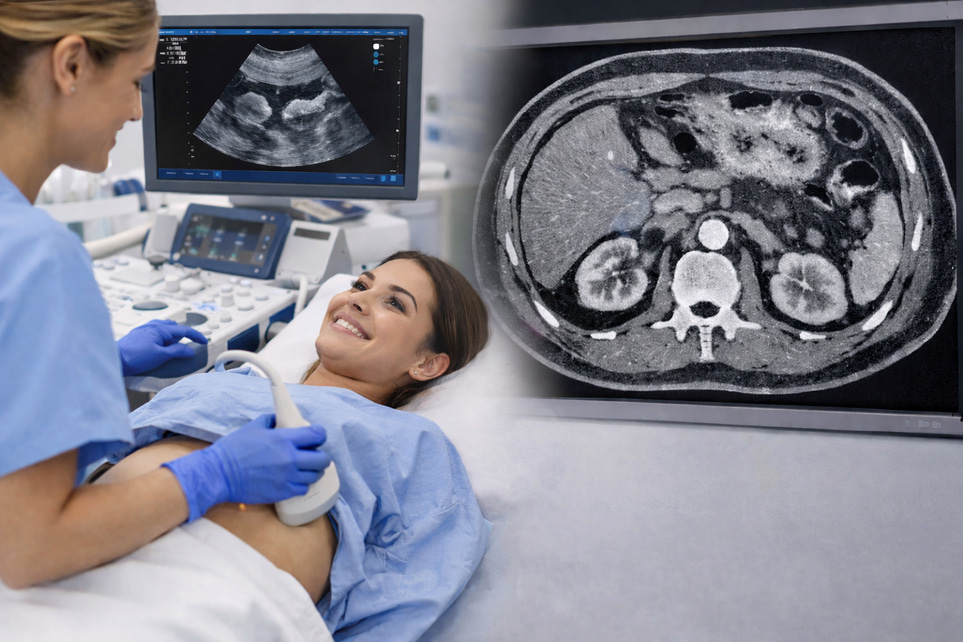
Computed tomography is an X-ray examination in which a rotating X-ray source takes many images from different angles. A computer then puts the images together into thin “slices” of the body, which allows the doctor to see organs, blood vessels, skeletons, and sometimes even small changes much more clearly than with regular plain X-rays.
That is why CT is often used when healthcare needs a quick and detailed answer. In the emergency room, the examination can help detect, for example, a brain hemorrhage, appendicitis, or internal injury after a fall. In a planned examination, it can be used to assess lungs, sinuses, blood vessels, or tumor disease.
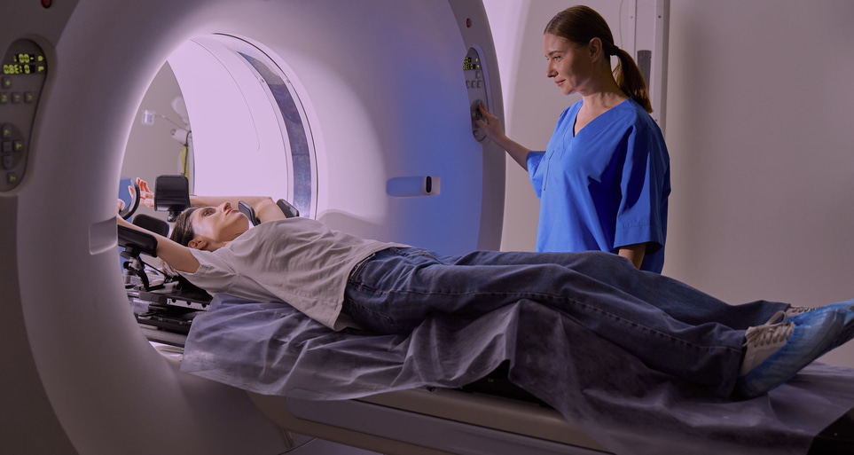
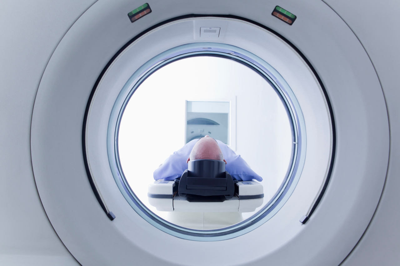
The camera itself looks like a short, wide ring with a couch that slides through the opening. Unlike MRI, the tunnel is short, and many people therefore experience CT as less cramped. The examination is not painful, but it is important to lie still so that the images are sharp.
How a CT examination is done in practice
Preparation depends on which part of the body is to be examined. Sometimes no special preparation is needed at all, while for other examinations you may be instructed not to eat for a few hours beforehand or to drink a liquid with contrast agent before the image is taken. The reception usually sends clear instructions in advance.
When you come to the X-ray, you will usually be asked to lie down on a couch. The X-ray nurse will help you into the correct position and then leave the room during the actual imaging, but will be in contact with you at all times via windows and speakers. You will often be asked to hold your breath for a few seconds, especially when examining the chest or abdomen, as breathing movements can otherwise blur the images.
Many CT examinations are quick. The actual imaging often only takes seconds to a few minutes, although the entire visit can be longer if you need to be prepared with a venous catheter, contrast medium or waiting time between different image series. For example, when examining kidney stones, the examination usually takes around ten minutes.
For children or adults who have difficulty lying still, the examination may sometimes need to be adjusted. In this case, the healthcare team plans for this in advance, as movement affects the image quality and in some cases can lead to the need to retake images.
CT with contrast medium – why it is sometimes necessary
In many examinations, contrast medium is used to increase the difference between different tissues in the images. This makes it easier to see, for example, blood vessels, inflammation, tumors or how urine passes through the kidneys and urinary tract. Contrast can be given into a blood vessel, swallowed as a liquid or, more rarely, given via the rectum, depending on what you want to examine.
When contrast medium is given into a vein in the arm, it is common to feel a quickly passing feeling of warmth in the body. Some people also get a metallic taste in the mouth or a feeling of having to urinate, even though this is not actually happening. This is unpleasant for some, but usually passes within a minute or so.
Most people do not experience serious side effects from iodine-based contrast medium, but mild reactions such as nausea, itching, redness or rash may occur. More pronounced allergic-like reactions are uncommon, and severe reactions are rare. Therefore, the staff always asks about previous contrast reactions, asthma, allergies and other diseases before the examination.
Contrast media, kidney function and metformin
Contrast media require extra care if you have impaired kidney function, as iodine-based contrast can rarely cause temporary kidney damage.
- Checking kidney function: Before the examination, check your kidney function often with a blood test (for example eGFR or creatinine).
- Pause of metformin: If you are taking metformin (a common medicine for type 2 diabetes) and have impaired kidney function, you need to take a break from the medicine in connection with the examination and for 48 hours afterwards.
- Resuming: Metformin is only restarted after a new blood test shows that kidney function is still stable. This is done because a temporary kidney effect from the contrast agent can otherwise increase the risk of serious side effects of the medication.
Radiation during computed tomography – how great is the risk?
CT uses ionizing radiation, and the radiation dose is higher than with regular X-rays. The Swedish Radiation Safety Authority states that the radiation dose to patients is significantly higher with computed tomography than with traditional X-ray examinations, and also states that a CT examination is often in the order of 1–10 mSv depending on which part of the body is examined and how the examination is carried out.
This does not mean that CT is dangerous in itself, but that the examination should be medically justified. A well-chosen CT can quickly provide crucial information that leads to the right treatment, and the benefit is usually much greater than the small long-term risk that the radiation entails. At the same time, radiology is actively working to keep the dose as low as possible and still obtain sufficiently good images.
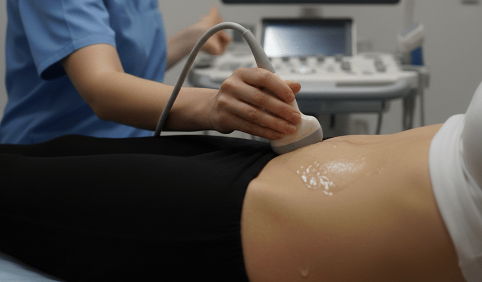
For example, a CT of the abdomen and pelvis is often associated with a higher radiation dose than many other X-ray examinations, while other CT protocols may be lower. The dose therefore varies greatly between different types of examinations and cannot be assessed fairly without knowing why the CT is being performed. This is also the reason why healthcare providers sometimes choose ultrasound or MRI instead, especially in children and pregnant women when medically possible.
If you have had many CT examinations before, it is reasonable to ask whether the new examination really adds anything. Such a question is not difficult for healthcare providers – it is wise. Radiation protection is based precisely on the fact that each examination should be justified and adapted to the patient's situation.
Frequently asked questions before and after a CT
One of the most common questions is whether the examination hurts. The answer is usually no. What you may feel is the needle prick if you get contrast in your blood, that the bed is hard or that you need to lie still in an unusual position for a short time.
Another common question concerns claustrophobia. Because the CT camera is short and open at both ends, many people usually cope with the examination better than they thought. If you know that you get very stressed in healthcare environments or confined spaces, it is still a good idea to let the staff know in advance, so that they can plan the visit calmly and clearly.
After the examination, most people can go home immediately or return to the ward. If you have received contrast medium, you can usually eat, drink and live as usual afterwards unless you have been given other instructions. The results are then reviewed by a radiologist, and the answer is passed on to the doctor who ordered the examination.
You should contact the healthcare provider afterwards if you experience a worsening rash, breathing difficulties or a clear feeling of illness after contrast medium, even if this is unusual. The same applies if you have received special instructions due to kidney function, a break from diabetes medication (such as metformin) or another concurrent illness.
CT provides answers to many acute and structural questions, but it does not show everything about how the body is doing on the inside. Fatigue, cardiovascular risk, anemia, diabetes, thyroid disorders or signs of low-grade inflammation are often captured better in blood tests than in X-rays. Therefore, imaging diagnostics and laboratory tests complement each other, rather than compete.