
Quick version
CT Lungs often only raise questions when something is already bothering you: a prolonged cough, shortness of breath that does not seem reasonable or an unexpected finding on a regular chest X-ray. For some, the examination becomes a way to investigate symptoms, for others it is about detecting changes early. CT Lungs can provide significantly more detailed information than a traditional X-ray and therefore be an important next step when you need clearer answers.
When can CT Lungs be a reasonable next step?
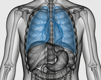
CT Lungs, or computed tomography of the lungs, is used when you want to see lung tissue, airways, pleura and structures in the chest in more detail than with regular X-rays. The examination may be appropriate for, for example, a long-term cough, repeated bloody cough, new shortness of breath, unclear chest pain or when a previous imaging finding needs to be assessed more closely.
CT Lungs, however, is not the right first choice for all respiratory problems. In many common conditions, clinical examination, blood tests, spirometry or a regular chest X-ray are sufficient. Computed tomography should be used when the benefits of the extra information are considered to outweigh the disadvantages, especially the radiation dose.
What can CT Lungs actually show?
One of the great strengths of CT Lungs is that the examination can detect small changes that are not visible on a regular X-ray. This includes lung nodules, i.e. small rounded changes in the lung tissue, which are sometimes completely benign but sometimes need to be followed up. CT can also show changes that are suspicious of tumors, enlarged lymph nodes, scarring, emphysema and signs of fibrosis, i.e. scar-like remodeling of the lung tissue.
The examination is also used for questions such as pneumonia that does not heal as expected, repeated infections, suspicion of bronchiectasis or more unusual interstitial lung diseases. It can also be central if you want to understand why a person is short of breath even though simpler investigations have not provided clear answers. Then CT can provide an anatomical explanation that complements lung function tests and blood tests.
In some cases, contrast agents are given in connection with the examination. This is done when you want to assess blood vessels, inflammatory changes or tumor spread more clearly. For example, in cases of suspected pulmonary embolism, a special CT technique is often used where contrast is needed to see blood clots in the vessels of the lungs.
CT Lungs as screening in smokers and former smokers
When CT is used for screening, it is not a regular diagnostic CT, but a low-dose CT. It is currently the only recommended screening method for lung cancer in adults with a clearly increased risk, primarily based on age and smoking habits. Recommendations state annual low-dose CT for people between 50 and 80 years of age who have smoked at least 20 pack-years and who still smoke or have quit within the last 15 years.
The aim is to find lung cancer at an earlier stage, when treatment can often be curative. Research and guidelines show that screening with low-dose CT can save lives if it is directed at people at increased risk. At the same time, it is important not to screen people outside the risk groups, as the disadvantages then risk being greater than the benefits.
For the individual, it is crucial to distinguish between screening and symptom assessment. If you have symptoms such as coughing up blood, weight loss or new shortness of breath, you should not think of "screening", but seek medical assessment for diagnostic assessment. Screening is intended for people without clear symptoms but with an increased risk profile.
If you find a spot, does it mean cancer?
Small changes in the lungs are common findings on CT, especially in people who have smoked, had infections or been examined for other reasons. Many nodules are scars, old inflammation residues or other benign changes. Therefore, a finding does not automatically mean that something dangerous has been discovered.
What determines the next step is, among other things, size, shape, density, growth over time and the person's risk factors. International guidelines are often used to support how incidentally discovered nodules should be followed up, for example whether a new CT scan should be performed after a few months or whether further investigation is needed immediately. However, the guidelines do not apply to everyone, for example not within formal screening programs or in people with known cancer or immunosuppression.
Another common finding is emphysema, which is a structural part of COPD where the alveoli have been damaged and their elasticity has decreased. CT can show such changes even before a person has put into words their decreased energy. This makes the examination valuable not only for finding cancer, but also for providing a clearer picture of other lung diseases that affect everyday life and work ability.
Limitations, radiation dose and when you should think twice
CT Lungs provides a lot of information, but it is not risk-free. Computed tomography involves a higher radiation dose than regular X-rays, and the Swedish Radiation Safety Authority states that computed tomography is generally significantly higher than traditional X-ray examinations. For chest CT, a typical effective dose is stated to be around 5 mSv, although the actual dose varies with the method, equipment and question.
Low-dose CT is used for screening, which means lower radiation than a diagnostic CT of the chest. Nevertheless, the benefit needs to be weighed against the risk, especially in younger people or when the probability of a clinically significant finding is low. Unnecessary CT examinations should be avoided, both to reduce radiation and to prevent chains of follow-ups after uncertain incidental findings.
There are also other limitations. CT can find changes that look suspicious but turn out to be benign, so-called false-positive findings. It can also miss some early or difficult-to-interpret changes. A normal CT response therefore does not rule out all causes of cough or shortness of breath, and the examination must always be interpreted together with symptoms, status, lung function and sometimes laboratory tests.
A wise use of CT Lungs is therefore not about examining as many people as possible, but about examining the right person at the right time. For some, the response is a safe confirmation that nothing serious is seen. For others, the finding becomes the starting point for early treatment, smoking cessation or further investigation before the disease has had time to cause more damage.