Quick version
Angiography is an X-ray examination used to examine blood vessels and detect conditions such as narrowing, blood clots and vascular malformations. Traditional angiography is done with a catheter that is inserted into the vessels, while modern CT angiography is a less invasive alternative where contrast medium is given through a vein in the arm.
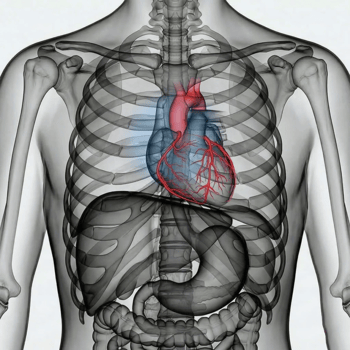
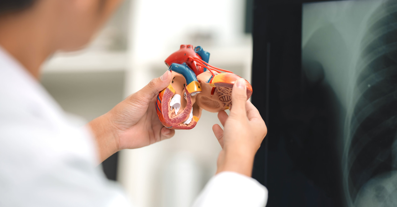
CT angiography is fast, gentle and provides very detailed images, which makes it particularly useful for examining, among other things, the coronary arteries of the heart. With us you can order CT coronary artery (CT coronary angiography) to detect changes in the blood vessels of the heart early.
The examinations are generally safe, require some preparation and are usually carried out without major discomfort. Most people can return to normal activities shortly afterwards, especially after CT angiography.
The examination is often used when there is suspicion of cardiovascular disease, stroke or other conditions where blood circulation may be affected. Today, there are also more modern variants where computed tomography is used, which makes the examination both faster and less invasive.
How angiography works
In a classic angiography, a thin catheter is inserted into a blood vessel, usually via the wrist or groin. Contrast medium is injected through the catheter, which makes the blood vessels clearly visible on X-ray images.
The doctor can then follow the passage of the contrast medium through the vessels in real time and detect:
- narrowings (atherosclerosis)
- blood clots
- aneurysms
- vascular malformations
The examination usually takes between 30 and 90 minutes and is performed under local anesthesia.
Different types of angiography
Angiography can be used to examine different parts of the body depending on symptoms and question.
- Coronary angiography (coronary artery X-ray) – examines the blood vessels of the heart in case of suspected angina or heart attack.
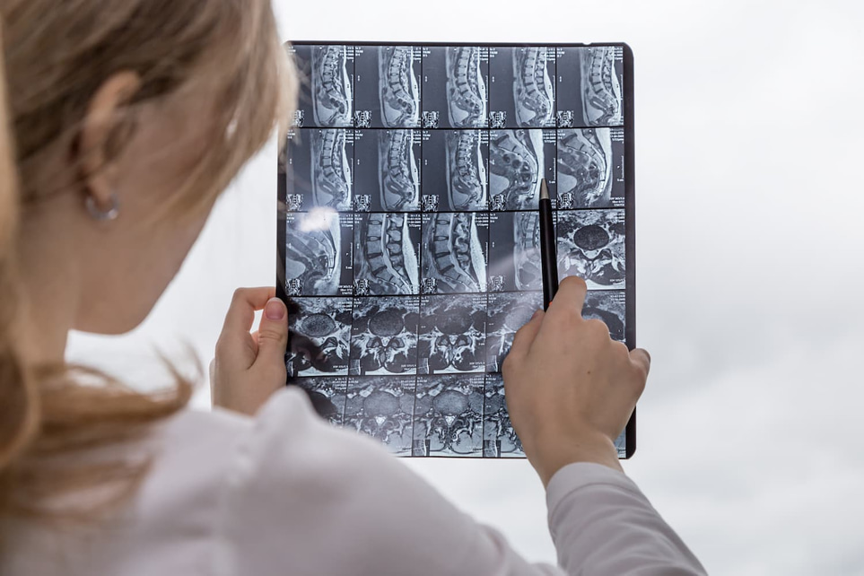
- Cerebral angiography – used to examine the blood vessels in the brain, for example in case of suspected aneurysm or stroke.
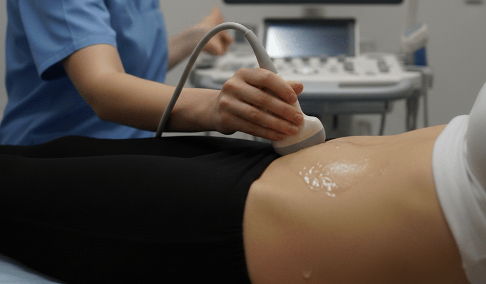
- Peripheral angiography – used to examine blood vessels in, for example, the legs, kidneys or gastrointestinal tract.
CT angiography – a less invasive alternative
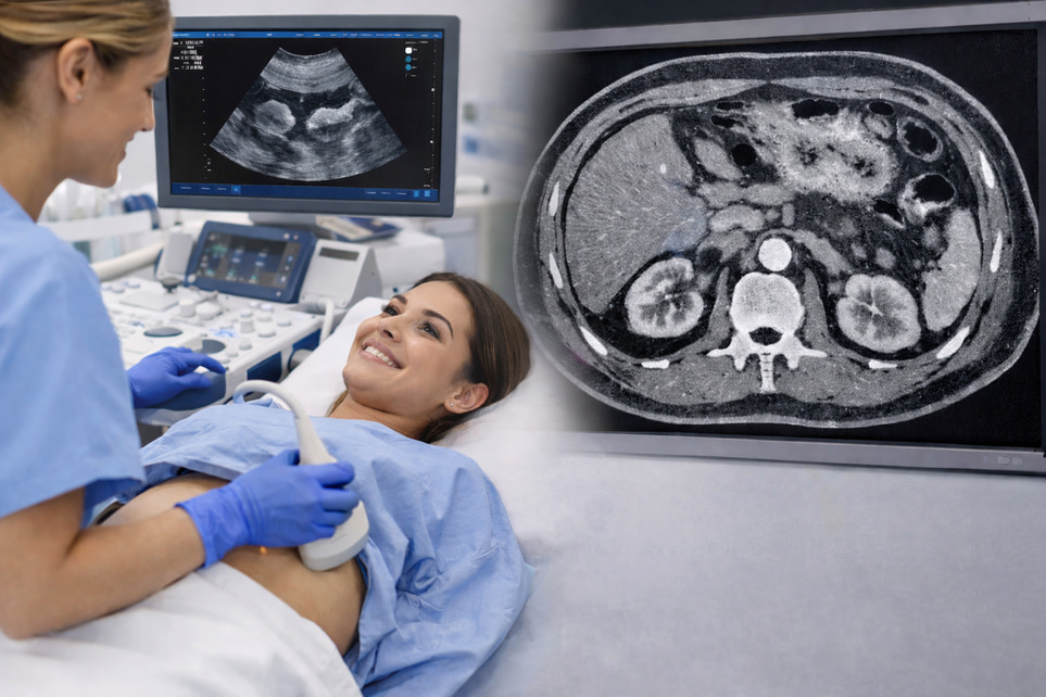
In many cases, computed tomography (CT) is used today instead of classical angiography. In a so-called CT angiography, contrast medium is given through a vein in the arm, usually in the crook of the arm, and images are taken quickly using a computerized tomography scanner that rotates around the body. The technique makes it possible to create very detailed cross-sectional images and three-dimensional reconstructions of the blood vessels.
Unlike traditional angiography, no catheter is needed to be inserted into the vascular system, which makes the examination less stressful for the patient. The imaging itself is quick, often in just a few minutes, although the entire visit may take a little longer depending on the preparations.
CT angiography is often used to examine, among other things:
- coronary arteries
- blood vessels in the lungs (in case of suspected pulmonary embolism)
- vessels in the brain
- the largest artery in the body (aorta)
The method has high diagnostic precision and can detect both narrowing, plaque formation and other structural changes in the vessels, often at an early stage.
Advantages of CT angiography:
- no catheter needs to be inserted into the vessels
- short examination time
- lower risk of complications
- can often be performed without hospitalization
- provides very detailed and three-dimensional images
CT coronary artery – An examination of the heart's vessels
With us you can order CT coronary artery (CT coronary angiography), a modern and gentle examination of the heart's blood vessels. The method provides very detailed images and can detect narrowing at an early stage.
The examination may be relevant for you who:
- have chest pain or suspected angina
- want to rule out narrowing of the coronary arteries
- have risk factors such as high blood pressure, high blood lipids or diabetes
- want to do a preventive check of the heart's vessels
CT coronary artery is a good alternative for many patients because it is both fast and less burdensome compared to traditional angiography.
Preparations for the examination
Before an angiography or CT angiography, you will always receive clear instructions from your healthcare provider. The preparations are intended to ensure that the examination can be carried out safely and provide the most reliable results possible.
Common preparations may include:
- blood tests to check kidney function, salt balance and blood status
- review of current medications
- information about any allergies to contrast media
- checking your pulse and blood pressure
In some cases, you may need to fast for a few hours before the examination, especially if contrast media is to be used. You will then receive specific instructions on how long you should abstain from food and drink.
Here is a more developed and in-depth version in the same style:How the examination is performed
How the examination is performed depends on the type of angiography used. Both classical angiography and CT angiography aim to visualize the blood vessels, but the methods differ in how they are performed.
With classical angiography:
- you will receive local anesthesia in the wrist or groin where the catheter will be inserted
- the doctor inserts a thin catheter into the blood vessel and navigates it to the area to be examined
- contrast agent is injected through the catheter, which may give a brief feeling of warmth in the body
- x-rays or ideosequences are taken in real time while the contrast agent passes through the vessels
You are awake throughout the examination and can communicate with the healthcare staff. In some cases, the doctor can treat a narrowing at the same time, for example by balloon dilation or stent insertion.
With CT angiography:
- contrast agent is given through a vein in your arm
- you lie on a table that is inserted into a CT scanner
- images are taken over a few minutes while you are asked to lie still and sometimes hold your breath for short periods
The examination is quick and painless. The entire visit usually takes less than an hour, although the imaging itself only takes a few minutes.
Is angiography dangerous?
Angiography is a safe and well-established examination that is routinely used in healthcare. X-ray radiation is used, but the dose is carefully adjusted to be as low as possible while maintaining image quality. The examination is always performed after a medical assessment where the benefits are considered to outweigh any risks.
With classical angiography, there is a small risk of complications linked to the catheter procedure, such as bleeding or bruising at the insertion site. With both classical angiography and CT angiography, contrast agents can rarely cause allergic reactions or affect kidney function, especially in people with already impaired kidney function.
How do you feel afterwards?
After a classical angiography, you may need to rest for a few hours. With CT angiography, you can usually return to normal activities immediately after the examination. Most people experience no discomfort afterwards and rarely need painkillers.