


MRI Lower Leg – Magnetic resonance imaging examination for pain, swelling or suspected injury in the lower leg
The lower leg consists of two bones – the shinbone (tibia) and the fibula – along with several muscles, tendons, blood vessels and nerves that extend down to the foot. The area is particularly vulnerable during sports, running, falls and strain injuries.
An MRI examination of the lower leg usually takes two projections – one in the frontal plane (from the front) and one in the lateral plane (profile). The examination provides a detailed view of bone tissue, tendons, muscles, ligaments and vascular structures. It is particularly useful for investigating suspected fractures, stress-related fractures, ligament or tendon ruptures and early signs of osteoarthritis or tissue wear.
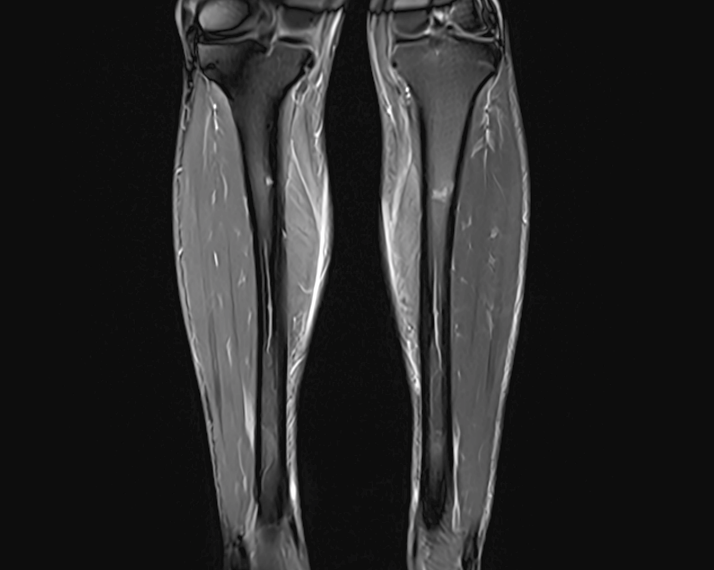
The examination takes approximately 30 minutes and is performed in a supine position. It is completely painless, radiation-free and provides high-resolution cross-sectional images of the various structures of the lower leg – even in difficult-to-assess cases where X-rays or ultrasounds do not provide sufficient information. The MRI scan is particularly valuable when X-rays have not been able to explain the symptoms, or when the injury is deep in the tissue.
When is an MRI scan of the lower leg recommended?
MRI of the lower leg is recommended for persistent or unexplained pain, suspected stress fractures, muscle injuries or inflammation. The scan is particularly common among athletes or people with strain-related problems that are not visible on regular X-rays.
- Long-lasting or recurring pain in the lower leg
- Suspected periostitis or compartment syndrome
- Swelling, bleeding or fluid accumulation after trauma
- Numbness, tingling or symptoms of nerve damage
- Suspected stress fractures in the tibia or fibula
- Infection or inflammation of the muscles or bone tissue
MRI is often used when the following conditions in the lower leg are suspected
- Stress fractures – small cracks in the bone that are often caused by overload
- Periostitis (medial tibial syndrome)
- Muscle ruptures or bleeding in the calf and shin muscles
- Chronic compartment syndrome – increased pressure in the muscle compartment
- Soft tissue or bone infections (osteomyelitis)
- Tumor or cyst in the bone or soft tissue of the lower leg
Book MRI Lower Leg – referral sent immediately
An MRI examination of the lower leg is an accurate and painless method for locating injuries, inflammations or structural changes. The examination takes approximately 15–25 minutes and is performed completely without radiation. We issue a referral immediately in connection with your booking, and you will receive a written specialist opinion within a few days.


