


Magnetic Resonance Imaging (MRI) for Foot Pain, Heel Pain or Suspected Foot-Related Injury
The foot is a complex structure consisting of 26 bones, 33 joints and over 100 tendons, muscles and ligaments – all working together to support the body’s weight and enable movement. This makes the foot particularly vulnerable to stress injuries, inflammation and degenerative changes that can affect both walking and quality of life.
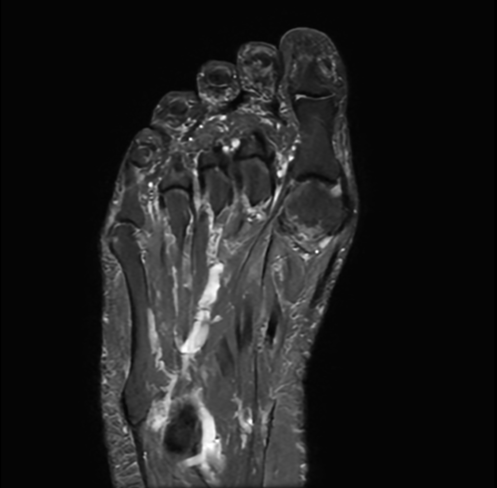
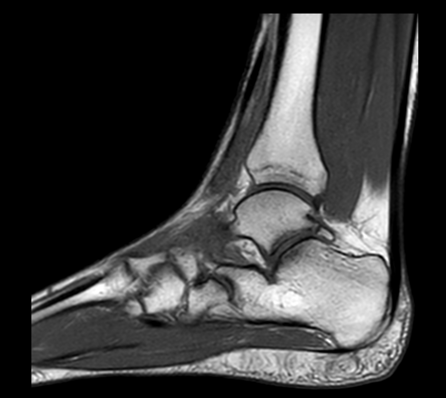
A magnetic resonance imaging (MRI) scan of the foot provides a high-resolution image of the internal structures of the foot – including the skeleton, articular cartilage, tendons, ligaments, joint capsules and soft tissues. The scan is completely radiation-free and provides a superior level of detail compared to regular X-rays or ultrasound, especially for complex pain or strain-related problems in the foot and heel.
MRI foot is particularly useful for long-term or difficult-to-explain foot pain, suspected stress fractures, tendon injuries, osteoarthritis, nerve compression or inflammation. The examination is painless, takes about 30 minutes, your referral is sent immediately after a review with one of our doctors and you will then receive a call for an MRI at one of the radiology units. Pain, stiffness or swelling in the foot can be caused by many different conditions – from overload and microfractures to rheumatic joint diseases or soft tissue tumors.
MRI is particularly valuable when other examinations have not provided clear answers or when injuries are suspected that are not visible on X-ray. You may consider a foot MRI if you experience:
- Persistent pain in the forefoot, midfoot or heel – especially with weight bearing or walking.
- Suspected heel spur or plantar fasciitis – where you want to assess inflammation and soft tissue involvement.
- Swelling, warmth or stiffness in the ankle or small joints – suspected arthritis or synovitis.
- Trauma to the foot – suspected fracture, ligament damage or tendon damage after a sprain or fall.
- Nerve pain, burning sensation or numbness – e.g. in case of suspected Morton's neuroma or tarsal tunnel syndrome.
- Changes in gait pattern or load-bearing capacity – where biomechanical disturbance or structural damage is suspected.
MRI provides a very clear image of both bone structure and soft tissues, which makes the examination particularly valuable for ensuring diagnosis prior to treatment or possible surgery.
MRI is often used when the following conditions in the foot are suspected
- Plantar fasciitis and heel spurs – inflammation of the tendon attachment under the foot, with or without skeletal overgrowth.
- Stress fractures – in the metatarsal bone, heel bone or metatarsal, which are often missed on conventional X-rays.
- Tendinitis and tendon injuries – e.g. in the tibialis posterior or peroneus tendons in cases of flat feet or overuse.
- Morton's neuroma – nerve compression between the metatarsal bones of the toes, often with burning pain in the forefoot.
- Osteoarthritis in the small joints of the foot – MRI shows early changes in cartilage and joint capsule that are not visible on X-ray.
- Arthritis or rheumatic joint diseases – such as RA, psoriatic arthritis or gout.
- Tumors or cystic changes – including ganglion, synovial cysts or other soft tissue tumors.
- Post-traumatic changes – in cases of persistent pain, stiffness or swelling after injury.
Book an MRI scan of the foot
An MRI of the foot is an advanced, non-invasive examination that provides important information about the cause of your problems. The examination is particularly valuable for symptoms that have not been answered by other methods or before a decision is made about treatment or surgery. An MRI of the foot takes about 30 minutes, is completely painless and does not involve radiation. The images are reviewed by experienced radiologists, and you will receive a written report within a few days.
Do you also have problems with your ankle? Since many injuries and diseases affect both the foot and ankle joint, it may be beneficial to perform an MRI of both areas at the same time. Contact us for advice on a suitable examination plan.


