Quick version
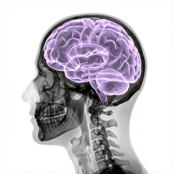
One of the most important examinations for suspected MS is an MRI of the brain. The disease causes inflammation in the nerve pathways of the brain and spinal cord, which can lead to damage to the myelin – the protective covering around the nerve fibers. These damages are often visible as so-called lesions or plaques on an MRI examination.
Doctors use MRI as a method to see if the changes have a pattern that is typical of MS and to assess whether the disease is spread in different parts of the nervous system and over time. At the same time, it is important to know that white spots on MRI do not automatically mean MS, since other conditions can also cause similar changes.
The diagnosis is therefore always based on a comprehensive assessment of symptoms, neurological examination, MRI findings and sometimes additional tests such as cerebrospinal fluid tests. After diagnosis, MRI is also used to monitor the development of the disease and evaluate the effect of treatment.
Common symptoms that may lead to an MS evaluation include numbness, vision problems, balance problems, muscle weakness, and severe fatigue. Early evaluation and treatment can reduce the risk of future injuries and disability.
Many people who experience symptoms such as numbness, visual impairment, dizziness or unusual fatigue describe the same feeling: What is happening in the nervous system? When the doctor suspects multiple sclerosis (MS), MR brain is often one of the most important examinations. The reason for this is that MS greatly affects the brain's white matter - the nerve pathways that function as the body's internal information cables - and that the changes can often be seen with an MRI.
What is the brain's white matter and why is it affected in MS?
Simply put, the brain consists of gray matter and white matter. The white matter mainly contains nerve fibers, called axons, which are covered with myelin. Myelin works much like the insulation around an electrical cable and allows nerve signals to be conducted quickly and efficiently. In MS, an inflammatory process occurs in the central nervous system where the immune system mistakenly attacks myelin and sometimes even the nerve fibers themselves. This leads to areas of demyelination, i.e. damage to the myelin, which can later develop into scar-like changes – what is called sclerosis.
That is why MS is often described as a demyelinating disease. When the myelin is damaged, signal transmission in the nerve pathways is impaired. Depending on where in the brain or spinal cord the inflammation is located, the symptoms can look very different.
Common symptoms can be:
numbness or tingling in the arms or legs
weakness or clumsiness
balance problems and dizziness
visual impairment or pain with eye movements, for example in optic neuritis
pronounced fatigue
cognitive problems, such as difficulty concentrating or slower thinking
MS does not only affect the white matter, but white matter lesions are still very important in diagnostics because they are often clearly visible with MRI. At the same time, it is known that even so-called normal-appearing white matter – white matter that looks relatively normal on a regular MRI – can be biologically affected in MS, which helps to explain why symptoms and functional impairment are sometimes greater than the number of visible lesions alone suggests.
What do MS lesions look like on a brain MRI?
A magnetic resonance imaging (MRI) scan of the brain is the most accurate non-invasive method for detecting typical MS changes in the brain. During the scan, the radiologist and neurologist look for lesions, sometimes called plaques, which are often visible as bright areas in the white matter on T2-weighted and FLAIR images. Typical MS lesions are often found in periventricular regions (around the brain's fluid-filled ventricles), juxtacortical or cortical areas (near or within the cerebral cortex), infratentorial regions (such as the brainstem or cerebellum), and the spinal cord.
What the doctor assesses is not only whether lesions are present, but also how they are distributed and whether they fit a typical MS pattern. This is crucial because white matter changes on an MRI do not automatically mean MS. Small vessel disease, migraine, aging and other inflammatory or autoimmune conditions can also cause changes in the white matter. Therefore, MRI must always be interpreted together with symptoms, neurological examination and sometimes cerebrospinal fluid testing.
A common question is: “Do white spots on MRI mean that I have MS?”
The answer is no. White spots can have several other causes. It is only when the appearance, location and clinical context of the lesions match that MRI becomes a strong support for the diagnosis. In some cases, contrast agents with gadolinium are used. If a lesion takes up contrast, it indicates that the inflammation is currently active or has recently been active, since the blood-brain barrier is then affected. Contrast thus helps primarily to show disease activity in time, while standard T2/FLAIR sequences show the total lesion burden over time. At the same time, development is underway towards more advanced non-contrast-based methods, but contrast can still be valuable in selected situations. When it comes to contrast agents, it is always the radiologist who decides whether this is considered necessary for the examination.
Why is brain MRI so important for diagnosing MS?
Making an MS diagnosis is based on being able to show that the disease is dissemination in space and time. This means that there must be signs that inflammation has affected different parts of the central nervous system and that this has not only happened on a single occasion. Brain MRI plays a key role here.
Dissemination in space can be supported by lesions in typical areas, for example:
periventricular
juxtacortical or cortical
infratentorial
spinal cord
optic nerve
Spread in time can be shown by:
both contrast-enhancing and non-contrast-enhancing lesions on the same examination
new lesions on a follow-up MRI compared with a previous MRI
sometimes by cerebrospinal fluid testing samples or other complementary findings according to current criteria
This is important because early diagnosis can allow for earlier treatment, which in turn can reduce the risk of new relapses, new lesions, and future disability.
Another common question is: “Can you have MS despite a normal brain MRI?”
In the early stages, the image can sometimes be difficult to interpret, and some people also need MRI of the spinal cord and in some cases optic nerve assessment, cerebrospinal fluid testing analysis or other tests. The diagnosis is therefore not made on a single image, but through a comprehensive neurological assessment.
Brain MRI for monitoring MS
Once the diagnosis has been made, MRI brain continues to be one of the most important tools for follow-up. The purpose is to see whether the disease is stable or whether new or growing lesions are appearing, even when the patient does not notice any clear new symptoms. In MS, so-called subclinical disease activity can occur, which means that the inflammation continues without causing an obvious relapse.
Follow-up MRI is used, among other things, to:
assess whether treatment slows down disease activity
detect new lesions early
assess whether treatment needs to be changed
monitor development over time in different forms of MS
It is also important to understand that MRI does not always reflect the entire disease picture. One person may feel relatively well despite multiple lesions, while another may have few but strategically placed changes that cause clear symptoms. Therefore, MRI is always combined with clinical assessment, functional status and the patient's own symptom picture.
When should you seek care?
If you have new neurological symptoms that do not go away – for example, unilateral numbness, loss of vision, double vision, balance problems or unexplained muscle weakness – you should seek care for an assessment. Just because you have these symptoms does not mean you have MS, but it is important to take them seriously. The sooner the right investigation is started, the better the chance of finding the cause and, if necessary, starting treatment early.
There is no blood test that can alone confirm or rule out MS, but blood tests are often important to rule out other conditions that can cause similar symptoms or affect the nervous system. Examples may include:
inflammatory or autoimmune conditions
infections
deficiencies or disorders that affect general health and nerve function
It is important to be aware that many symptoms that raise concerns about neurological disease can be related to more common and treatable abnormalities in the body's values.
MS is a complex disease where inflammation often attacks the white matter of the brain and creates lesions that can be detected with MR brain. MRI helps the doctor understand where in the nervous system the damage is located, whether the disease is active and how it develops over time. For those living with symptoms, this can provide both clearer answers and better opportunities for the right treatment.