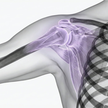
An MRI examination of the shoulder examines the structures that are usually behind pain and impaired function. Since many shoulder problems are located in the soft tissues such as tendons, muscles and cartilage, MRI is a relatively accurate method for identifying the cause.
Unlike a regular plain X-ray that mainly shows the skeleton, MRI can clearly visualize the rotator cuff, the cartilage of the joint and other important structures in the shoulder. This makes the examination particularly valuable in cases such as long-term pain, suspected tendon injuries or when mobility in the shoulder is impaired without a clear explanation.
The shoulder is also one of the most mobile joints in the body, which places high demands on the interaction between muscles and tendons. When something in this system is affected, it can cause symptoms that are not always visible on an X-ray but that often appear on an MRI.
Common symptoms of shoulder problems
Many people who undergo an MRI of the shoulder have had problems for a long time. The pain often starts diffusely and can be difficult to pinpoint, but tends to gradually worsen. In other cases, symptoms appear more suddenly, for example after lifting, a training load or a jerk in the arm.
Shoulder problems often affect both mobility and strength, and can make everyday activities such as getting dressed, lifting things or sleeping undisturbed significantly more difficult. It is also common for the pain to vary over time, with periods of worsening with load.
- Pain when lifting the arm, especially outward or above shoulder height.
- Nocturnal pain, often when lying on the affected side.
- Feeling of weakness or that the arm “doesn’t really carry”.
- Stiffness or limited mobility in the shoulder joint.
- Pain with specific movements or loads, for example during exercise.
In some cases, the symptoms may be due to irritation or wear and tear in the tendons and joints, while in other cases it may be a more obvious tendon injury. When the symptoms do not improve over time, or when the function of the shoulder is clearly impaired, an MRI examination can be an important step in understanding the cause and moving forward with the right treatment.
This is what an MRI of the shoulder can show
An MRI of the shoulder provides a detailed image of both joint structures and soft tissues, making it possible to identify changes that are not visible with other imaging methods. Often it is a combination of several findings – rather than a single injury – that together explain the symptoms.
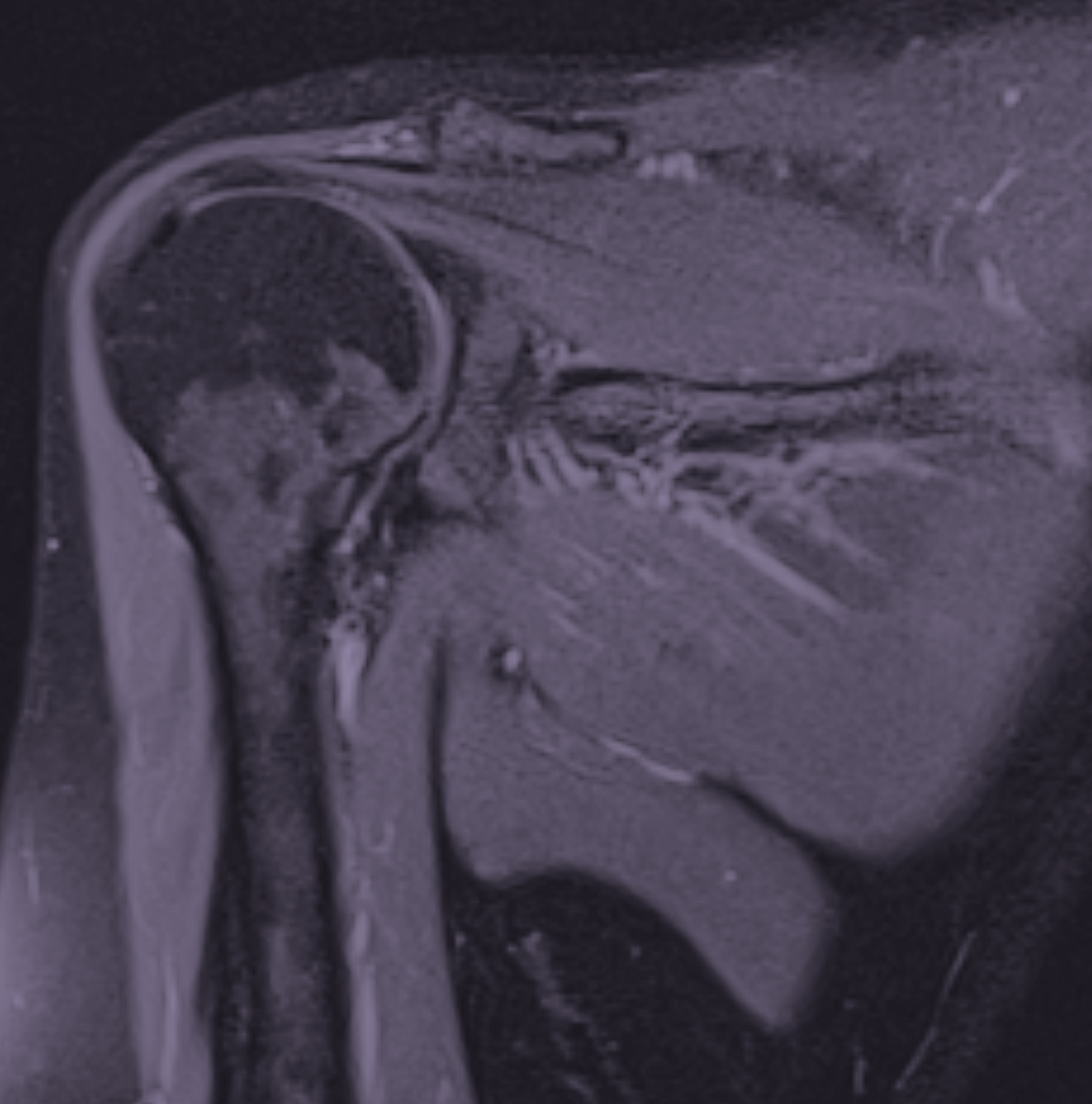
Wear and tear changes in the joint
Degenerative changes are common, especially in the acromioclavicular joint (AC joint) on top of the shoulder. These changes include cartilage damage, joint space narrowing, and bone spurs (osteophytes). In some cases, osteophytes from the acromion can contribute to reduced space in the subacromial space, which can affect the rotator cuff tendons and cause pain during movement.
Changes in the rotator cuff
The rotator cuff consists of four tendons that together stabilize the shoulder joint and enable movement. On MRI, it is common to see tendinosis, which means degenerative changes in the tendon tissue with reduced structure and strength. This is often related to long-term strain, age, or reduced circulation in the tendon.
MRI can also show partial ruptures, where parts of the tendon fibers are damaged without the entire tendon being severed. The tendon most often affected is the supraspinatus tendon. Typical findings may include thinning of the tendon, irregular signal, or that parts of the tendon fibers can no longer be followed in their normal stretch. In some cases, retraction is also seen, which means that the tendon has pulled slightly from its attachment. This type of change often indicates a more chronic rather than acute injury.
Mechanical impact on the shoulder
MRI can also show if there are cramped conditions in the shoulder where structures press against each other. This can occur when the shape of the skeleton or accumulations reduce the space for the tendons, which over time can lead to irritation and damage.
Muscles and biceps tendon
In addition to tendons, the quality and structure of the muscles are also assessed. MRI can show whether the muscles are unaffected or if there are signs of long-term strain. The biceps tendon is also analyzed to ensure that it is intact and lies correctly.
How to interpret the findings
It is important to understand that many changes seen on MRI can also occur in people without symptoms. Mild degenerative changes and tendinosis are common, especially with increasing age, and do not always have to be the cause of the symptoms.
The interpretation of an MRI is always done in relation to the clinical picture. Your symptoms, your medical history and the question that forms the basis of the examination are of utmost importance. A holistic assessment is crucial to determine which findings are relevant and whether they require further action.
Depending on what the examination shows, in some cases further investigation or referral to a specialist, such as an orthopedist, may be necessary. At the same time, it is common for treatment to be primarily conservative, with a focus on, for example, physiotherapy, adapted training and weight-bearing.
MRI therefore often serves as an important basis for decision-making, both to rule out more extensive injuries and to guide the correct treatment based on your specific situation.
When is it worth doing an MRI of the shoulder?
An MRI of the shoulder can be an important next step when the symptoms do not go away or when the cause is unclear. For many, a private examination means that you get answers faster and can proceed with the right treatment, without having to wait in long healthcare queues.
- Pain that has been present for several months.
- No improvement despite physiotherapy or rest.
- Clear weakness or difficulty lifting the arm.
- Suspected tendon damage after strain or injury.
By identifying the cause of your symptoms early, the possibility of initiating the right treatment increases and the problems do not worsen over time.
If you have persistent pain or impaired function, an MRI can be an important step in understanding the cause and moving on to the right treatment. With us you can order a MRI of the shoulder we will send a referral after consultation and you will always receive a medical assessment based on your results.