Quick version
A slightly heterogeneous thyroid gland with small cystic changes is a common ultrasound finding and may be a sign of a previous thyroiditis, also called thyroiditis. If no tumor-like changes or signs of active inflammation are seen, it is often a benign finding. To get a complete picture, the ultrasound result needs to be assessed together with blood tests, symptoms and any previous medical history.
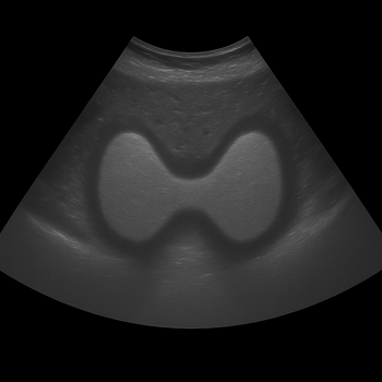
Ultrasound is a valuable tool for mapping the structure of the thyroid gland and can provide important clues about both previous and ongoing disease processes.
What is thyroiditis?
Thyroiditis is a collective term for inflammation of the thyroid gland. The inflammation can be both temporary and long-term and can occur for several different reasons. The most common forms are:
Hashimoto's thyroiditis (autoimmune thyroiditis)
Subacute thyroiditis (often after a viral infection)
Silent or painless thyroiditis
Postpartum thyroiditis (after pregnancy)
Depending on the type and stage of the disease, thyroiditis can affect the production of thyroid hormones and lead to both hyper- and hypofunction. Here you can read more about symtom vid tyreoidit and its different forms.
What does thyroiditis look like on ultrasound?
A healthy thyroid gland normally has a smooth structure and a homogeneous appearance on ultrasound. During or after an inflammation, the tissue can change.
Common ultrasound findings in thyroiditis are:
Heterogeneous (uneven) tissue structure
Small cystic changes (small fluid-filled sacs)
Areas with altered echogenicity
Enlarged or sometimes smaller thyroid gland
Altered blood flow in the gland
In some cases, ultraljud av sköldkörteln can show a slightly heterogeneous thyroid gland with small cysts without any active inflammation continuing. This may be a sign that the gland has previously been inflamed and left structural changes.
What does a slightly heterogeneous thyroid gland mean?
The term "heterogeneous" means that the tissue does not look completely smooth on the ultrasound image. It is a relatively common finding and does not necessarily mean that there is an ongoing disease.
A slightly heterogeneous structure can be seen if:
you have previously had thyroiditis
there is an early autoimmune thyroid disease
there are age-related changes
mild chronic inflammatory changes have occurred
The finding always needs to be assessed together with blood tests and any symptoms, as the ultrasound shows what the gland looks like, not how it works.
Small cysts in the thyroid gland – are they dangerous?
Small cysts in the thyroid gland are common and usually completely benign. They consist of fluid-filled cavities that can occur in connection with normal tissue changes or after a previous inflammation.
Smaller cysts without suspicious characteristics usually do not require treatment or further investigation.
When is there cause for concern?
Ultrasound is used, among other things, to detect nodules that may need to be examined more closely. Certain features may raise suspicion of tumor disease, for example:
Irregular edges
Microcalcifications
Pronounced low echogenicity
Increased vascular supply in suspicious areas
Enlarged lymph nodes in the neck
If no such findings are found, the risk of cancer is usually very low.
Do you need to take more tests?
Yes, if the ultrasound shows changes that may indicate previous thyroiditis, the doctor can supplement with blood tests to assess the function of the thyroid gland.
Common tests that are then performed are:
These tests can help determine if there is an ongoing autoimmune process or impact on hormone production.