MRI Forearm – Magnetic resonance imaging examination for pain, nerve damage or soft tissue injuries in the forearm
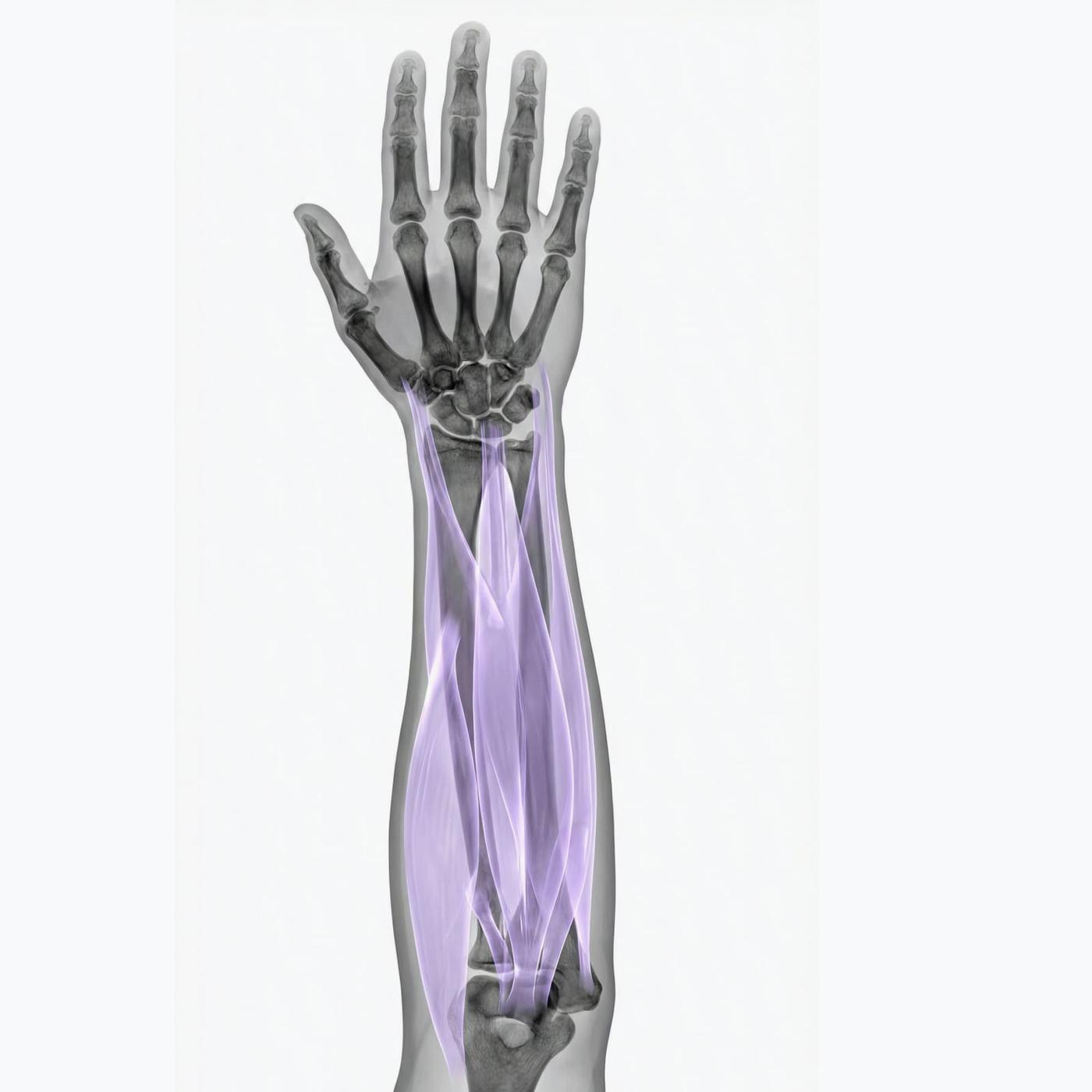
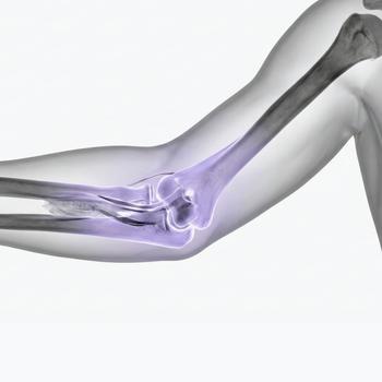
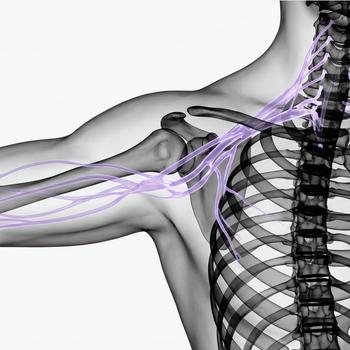
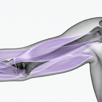
The forearm consists of two long bones – the radius and the ulna – as well as a complex network of muscles, tendons, blood vessels and nerves. The area is often exposed to stress injuries, inflammation or nerve compression that can affect both mobility and function in the hand and wrist.
An MRI examination of the forearm provides a detailed image of both the skeleton and soft tissues and is particularly useful in cases of long-term pain, suspected nerve damage (e.g. ulnar nerve syndrome), tendon problems or after trauma. The MRI can identify injuries that are not visible on regular X-rays, completely without radiation.
When is an MRI examination of the forearm recommended?
An MRI of the forearm is an important tool for pain that does not go away, or when damage to tendons, muscles or nerves is suspected. It is also used to map inflammation or cystic changes in connection with repeated loads or sports injuries.
- Long-term pain or tenderness in the forearm or wrist.
- Numbness, tingling or loss of sensation in the hand or fingers.
- Suspected nerve compression – e.g. ulnar nerve syndrome or radial nerve involvement.
- Tendon injuries, inflammation or muscle changes.
- After trauma – suspected soft tissue injury or stress fracture.
- Swelling, cysts or other structural changes.
MRI is often used when the following conditions are suspected in the forearm
- Tendinitis – inflammation of tendons after overexertion.
- Nerve compression – e.g. ulnar neuropathy or radial entrapment.
- Muscle injuries or bleeding after sports or trauma.
- Ganglion or other soft tissue tumors.
- Stress fracture in the radius or ulna that is not visible on X-ray.
- Myositis – suspected muscle inflammation or infection.
Book an MRI of the forearm – safe and accurate examination
An MRI examination of the forearm is an accurate, painless and radiation-free method for identifying the cause of your symptoms. The examination takes approximately 15–25 minutes and we issue a referral immediately in connection with the order. The images are reviewed by a specialist and you will receive a report within a few days.

