MRI Skull Base - for nerve damage, tumor or suspected change in the skull base area
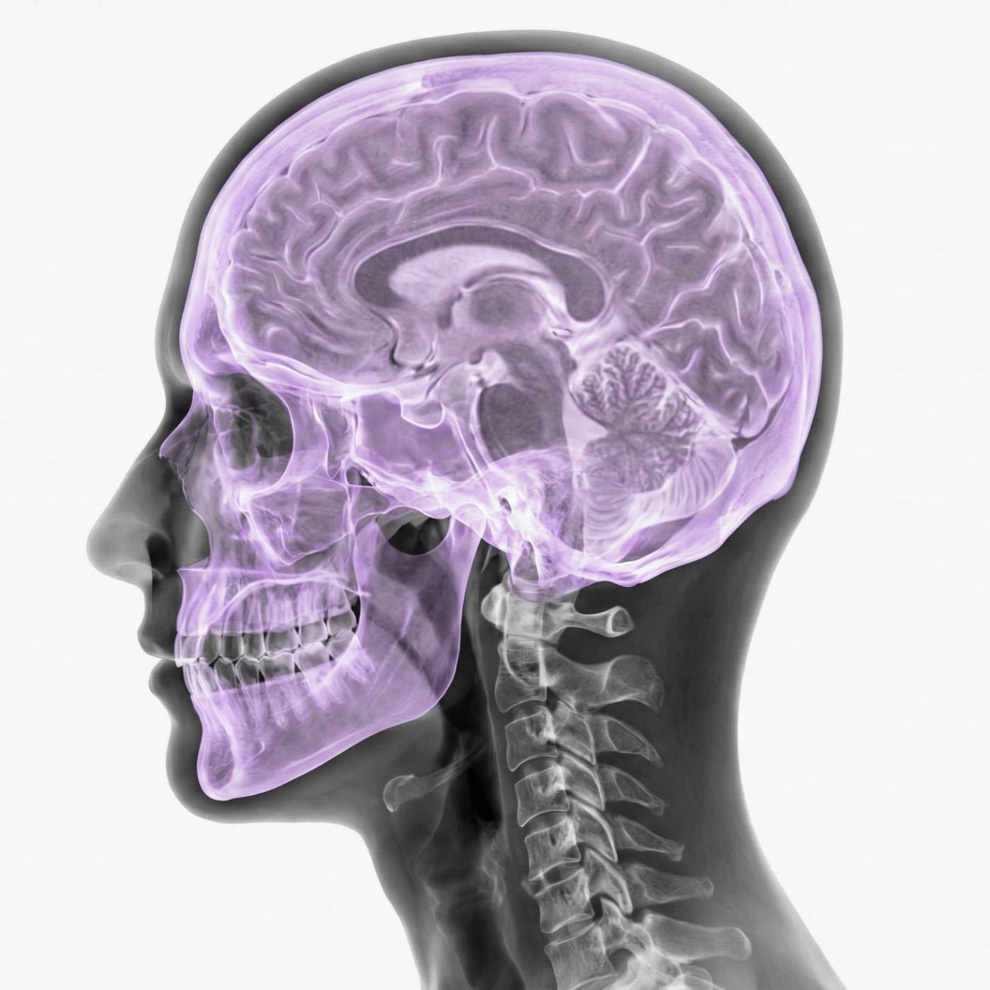
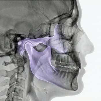
The skull base is an anatomically complex structure that forms the boundary between the brain and the face/neck region. Several important nerves, blood vessels and channels pass through the skull base, making the area susceptible to both benign and serious diseases. If you experience symptoms such as unilateral facial paralysis, numbness, hearing loss, vision changes or unclear pain in the face or neck, an MRI scan of the skull base can be crucial in identifying the cause.
MRI Skull Base is a high-resolution, radiation-free examination that maps both bone structures, soft tissues and nerve pathways. It is often used as an in-depth investigation when a tumor, nerve compression, vascular malformation or inflammatory processes in the area are suspected. The examination is also valuable before planned surgery or when following up on previously known changes.
When is an MRI of the skull base an important examination?
Because the skull base is a central structure through which several important nerves, blood vessels and cavities pass, even small changes can give rise to significant symptoms. MRI of the skull base is usually ordered for unclear neurological problems or suspicion of a tumor, infection or vascular change affecting the tissues of the skull base. Below are listed common medical indications for performing the examination.
- Suspected tumor or metastasis in the skull base.
- Nerve involvement, facial paralysis or numbness.
- Dizziness, balance disorder or tinnitus where other investigations have been without findings.
- Pain in the face or behind the eyes that is not explained by another cause.
- Chronic infections or suspicion of inflammation in the skull base region.
- Mapping of vascular malformations, fistulas or bone involvement.
What can an MRI scan of the skull base show?
- Changes in the bones of the skull base, e.g. erosion or infiltration.
- Pressure on or interruption of nerve pathways – especially cranial nerves.
- Tumors such as meningioma, schwannoma or chordoma.
- Inflammatory changes, abscess or fibrosis.
- Vascular malformations or venous stasis in nearby structures.


