MRI Orbita – Magnetic resonance imaging examination for eye problems, vision impairment or suspected changes in the eye socket
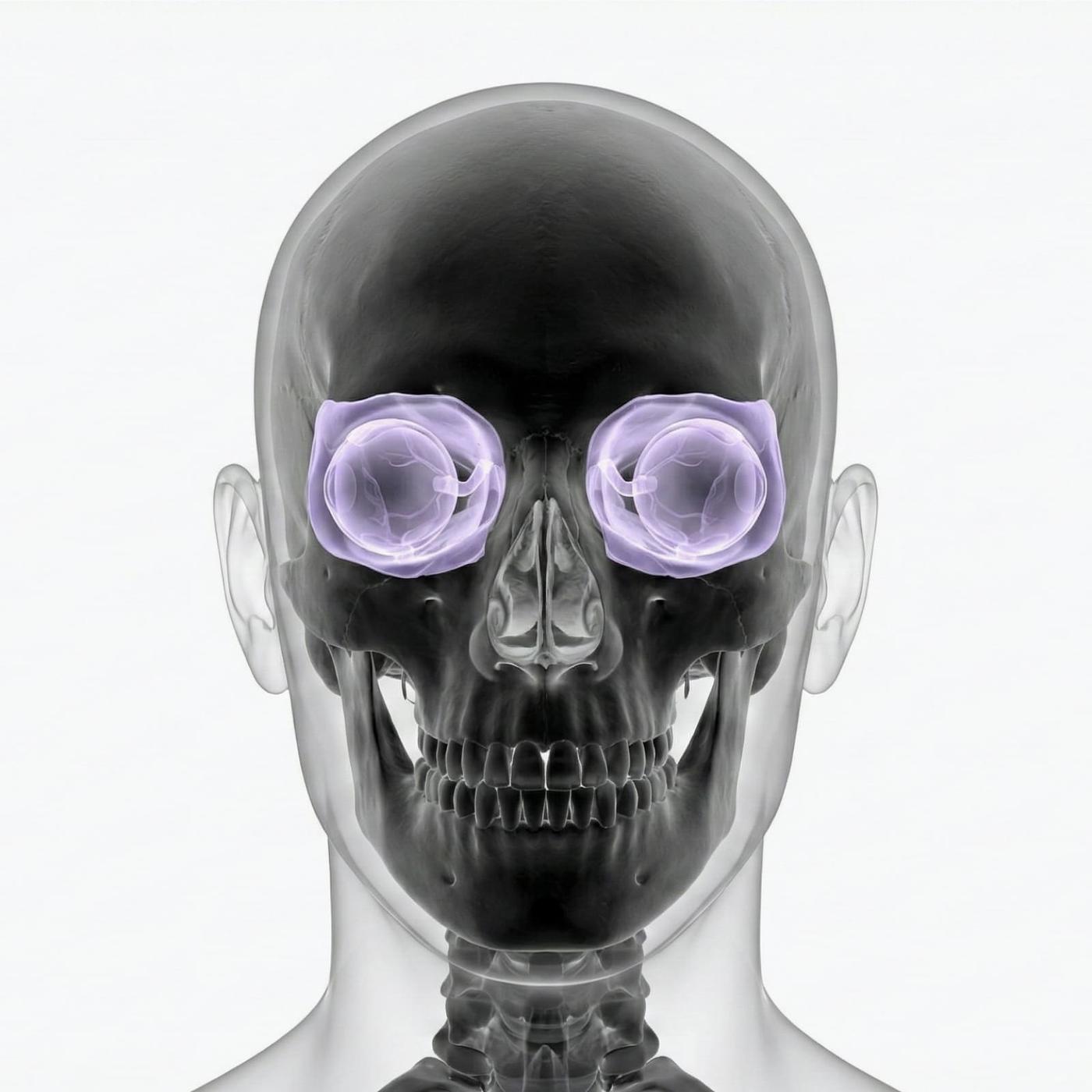
Have you suffered from vision impairment, pressure behind the eye, double vision or swelling in the eye region? Then an MRI examination of the orbit can be crucial in finding out the cause. The examination provides high-resolution images of the eyeballs, optic nerve, eye muscles, fatty tissue and the internal structures in the eye sockets – completely without radiation. It is used to identify tumors, inflammation, nerve damage or other changes that are not visible during regular eye examinations.
MRI examination of the eye sockets is often used when regular eye examinations are not enough to identify the underlying causes of symptoms. MRI orbita is recommended when tumors, inflammation, bleeding, nerve damage, changes in the visual pathway or injuries after trauma are suspected.
When is an MRI of the orbit recommended?
MRI of the orbit is used to assess structures in and around the eye that cannot be seen with external examination or regular X-rays. It is particularly useful in neurological or inflammatory conditions that affect eye movements or vision.
- Visual impairment without a clear cause (visual impairment, blurred vision, double vision).
- Suspected optic neuritis (inflammation of the optic nerve).
- Feeling of pressure, swelling or pain behind the eye.
- Protruding eye (proptosis) or asymmetry.
- Suspected tumor in the orbit, such as meningioma, lymphoma or glioma.
- Investigation of trauma to the eye region.
MRI is often used when the following conditions in the eye socket are suspected
- Optic neuritis or other diseases of the optic nerve.
- Orbital pseudotumor (inflammatory mass in the eye socket).
- Endocrine ophthalmopathy (e.g. Graves' disease).
- Tumors in the orbit, eyeball or optic nerve canal.
- Bleeding, infection or abscess in the orbit.
- Injuries after trauma or fracture of the orbital wall.
Book an MRI of the Orbit – Get a referral immediately
The examination takes approximately 20–30 minutes and is completely painless. You lie still in the magnetic resonance imaging camera while high-resolution images are taken of the eye sockets and nerve structures. A referral is not needed – we issue it directly upon booking. The answer will come from a specialist doctor within a few days.

