


MRI of the Pelvis and hip Joint
Do you experience pain in your hip or pelvic area? You can now book a private MRI scan without a referral and get an appointment within just a few days. An MRI is recommended if you’ve had persistent pain, stiffness, or limping that hasn’t improved with rest or treatment.
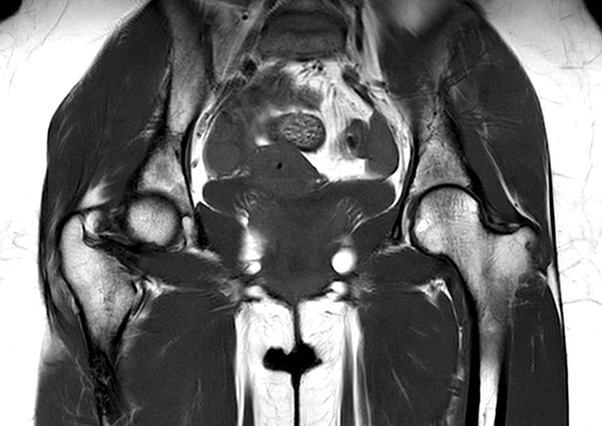
MRI of the pelvis and hip provides high-resolution images of bones, cartilage, tendons, bursae, and surrounding soft tissues – entirely without radiation. The examination is completely painless and can identify everything from early signs of osteoarthritis to inflammation or soft tissue injury.
When is an MRI recommended for hip or pelvic pain?
Pain in the hip or pelvis can have many causes – from overuse and muscle injuries to rheumatic diseases or nerve compression.
In many cases, X-ray or ultrasound does not provide sufficient information, especially when early changes in cartilage, bone, or soft tissue are suspected.
MRI is then the most accurate method for identifying the underlying issue. It may be beneficial to schedule an MRI of the pelvis and hips if you experience any of the following symptoms:
- Deep, persistent pain in the groin, hip, or buttock – that does not improve with movement, rest, or physiotherapy.
- Limping or reduced hip mobility – especially if it affects walking, climbing stairs, or hip rotation.
- Night-time pain in the hip or pelvis – that disrupts sleep and may indicate inflammation.
- Sudden pain after a fall, sports injury, or accident – where muscle tear, stress fracture, or labral injury is suspected.
- Radiating pain to the thigh, lower back, or pelvic floor – which may be due to nerve involvement in the lumbar spine or pelvic region.
- Swelling, stiffness, or instability in the hip joint – that cannot be explained by other investigations.
If you’ve had previous imaging or treatment without a clear diagnosis, an MRI may provide crucial information to guide your next steps.
MRI is commonly used to assess the following conditions in the hip and pelvis
- Hip osteoarthritis – degeneration of joint cartilage causing pain, stiffness, and reduced mobility. MRI can detect early cartilage changes before they appear on X-ray.
- Labral tear (wear or rupture of the acetabular labrum) – a common cause of deep groin pain in younger, active individuals.
- Trochanteric bursitis or tendon inflammation – common in women over 40. MRI confirms the diagnosis and excludes other pathologies.
- Rheumatic joint disease – e.g., sacroiliitis in ankylosing spondylitis or joint involvement in rheumatoid arthritis. MRI can visualize active inflammation in bone and joint capsule.
- Stress fractures – common in runners and athletes, especially in the pelvis or femoral neck. Often only visible on MRI.
- Snapping hip syndrome and impingement – conditions where muscles or tendons catch or rub, sometimes causing inflammation and pain.
- Pelvic instability or symphysis dysfunction – e.g., after pregnancy, trauma, or hypermobility. MRI can provide important information about ligaments and joints in the pelvic ring.
- Tumors, cysts, or other abnormalities – that cannot be sufficiently evaluated with standard imaging techniques.
Book an MRI of the Hip or Pelvis
An MRI of the hip or pelvis is a powerful diagnostic tool that provides detailed insights into bones, joint surfaces, tendons, cartilage, bursae, and surrounding soft tissues.
This examination is especially valuable in cases of complex symptoms, diffuse pain, or when there is a need to rule out more serious conditions. The scan takes approximately 30 minutes, is completely painless, and is performed without any exposure to radiation.


