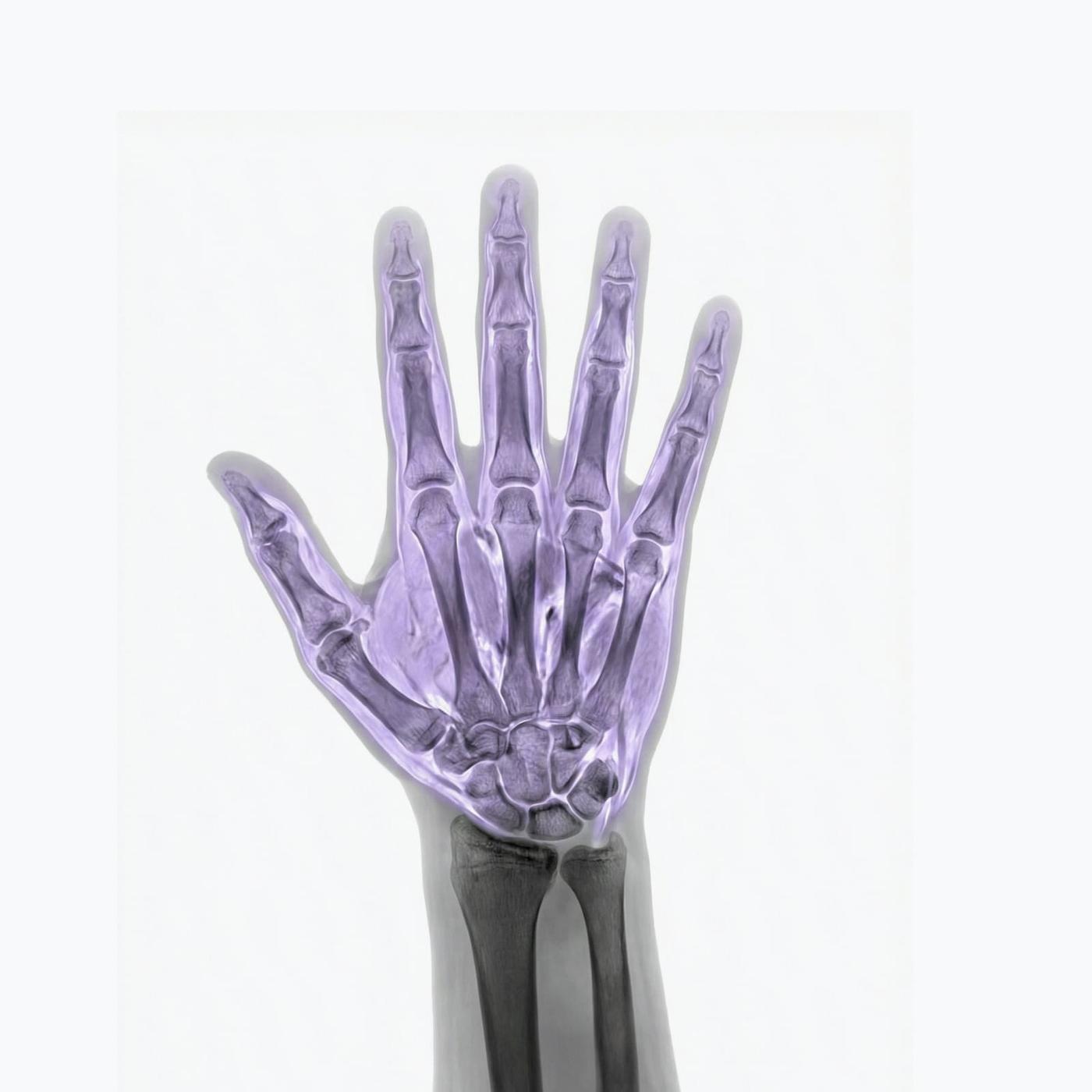
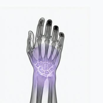
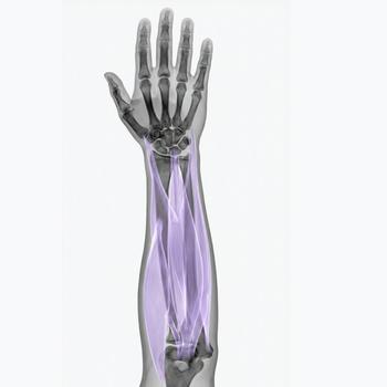
The hand is composed of numerous small bones, joints, tendons, muscles, and nerves that together enable fine motor control, grip strength, and force transmission. This intricate anatomy means that even minor injuries or inflammation can significantly impact daily function and work capacity.
With a magnetic resonance imaging (MRI) scan of the hand, highly detailed images of the internal structures can be obtained – including bones, soft tissues, cartilage, tendons, blood vessels, and nerves. The examination is completely radiation-free and often provides more accurate diagnostic information than both X-ray and ultrasound for many conditions affecting the bones and soft tissues of the hand.
Hand MRI is especially useful for complex or unclear pain, post-traumatic evaluation, suspected tissue damage, or in the assessment of rheumatic or inflammatory diseases. The procedure is painless, takes approximately 20–30 minutes, and requires no referral – you can book directly.
When is an MRI of the Hand Recommended?
Pain or stiffness in the hand can be caused by a variety of conditions – ranging from minor fractures and tendon injuries to joint inflammation, nerve entrapment, or tumor-like lesions. MRI is primarily used when there is a need to visualize structures not adequately seen on conventional X-ray or ultrasound. You may consider a hand MRI if you are experiencing:
- Localized painful lumps or swelling in the hand – suggestive of a ganglion cyst, tumor, or other mass.
- Reduced finger function or inability to flex or extend – which may indicate tendon injury or rupture.
- Persistent, unexplained hand pain – especially when other examinations have not provided a diagnosis.
- Suspected fracture or stress fracture – not visible on standard X-ray.
- Inflammatory conditions – such as rheumatoid arthritis or psoriatic arthritis involving small joints.
- Nerve-related symptoms in one or more fingers – such as numbness, tingling, or weakness.
MRI is often the best choice when a definitive diagnosis is needed or when evaluating the extent of an injury prior to treatment or surgery.
Common Indications for Hand MRI
- Tendon ruptures and soft tissue injuries – e.g., after trauma or suspected tears in flexor or extensor tendons.
- Ganglion or other cystic lesions – MRI determines size, location, and relation to adjacent structures.
- Tumors or tumor-like processes – including both benign and malignant lesions in bone or soft tissue.
- Joint inflammation (synovitis) – commonly seen in rheumatic diseases; MRI reveals joint fluid, swelling, and tissue changes.
- Arthritis or erosions – especially in small joints affected by RA, psoriatic arthritis, or SLE.
- Fractures – including microfractures or stress fractures that may not be visible on standard radiographs.
- Vascular abnormalities – such as vessel malformations or hemangiomas within soft tissues.
- Post-traumatic changes or functional impairment – MRI provides a complete evaluation after injury.
Book an MRI of the Hand
Hand MRI is a highly advanced imaging modality that enables precise and reliable diagnosis of hand conditions. It is used both for acute injuries and for chronic, unexplained symptoms that have not been clarified by previous evaluations. The scan takes about 20–30 minutes, is entirely painless, and does not involve radiation


