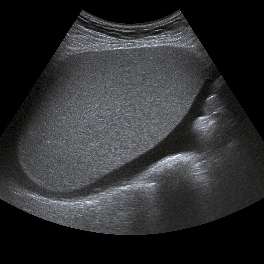
A spleen ultrasound is used to examine the size, shape, tissue structure and blood flow of the spleen. The examination is performed by a specialist in radiology and provides detailed images in real time that can show signs of enlargement, cysts, infarctions or other changes. Spleen ultrasound is often used to investigate the cause of abdominal pain, infection, anemia, suspected enlarged spleen (splenomegaly) or as part of an investigation of liver and blood diseases.
Spleen ultrasound – for pain, enlargement or suspected impact on the blood picture
The examination is recommended for symptoms such as pain or pressure under the left rib cage, repeated infections, fatigue, anemia or suspected enlarged spleen. Ultrasound can assess both the size and structure of the spleen, which is important if a blood disease, infection, trauma or liver disease is suspected. It is also used to follow up on conditions following infections such as mononucleosis (glandular fever) or other viral diseases that can affect the spleen.
Unlike MRI and CT, which are used for more detailed mapping of the internal structure or vascular supply of organs, ultrasound is the first-line method for assessing spleen size, blood flow, and superficial changes. The examination is quick, accessible, and completely radiation-free.
Common symptoms and questions
- Pain or pressure under the left rib cage.
- Suspected enlarged spleen (splenomegaly).
- Recurrent infections or anemia.
- Check for glandular fever or other infection affecting the spleen.
- Suspected cysts, infarctions or tumor changes in the spleen.
- Follow-up after trauma to the abdomen or suspected spleen injury.
Conditions that can be detected with ultrasound of the spleen
- Splenomegaly – enlarged spleen as a result of infection, liver disease or blood disease.
- Splenic infarction – reduced blood flow to parts of the spleen.
- Cysts, abscesses or other fluid-filled changes.
- Tumor changes or metastases in the spleen.
- Splenic injuries or hematomas after trauma.
- Altered blood flow in portal hypertension or liver disease.
How an ultrasound of the spleen is performed
The examination is performed while you lie on your back or on your right side. A gel is applied to the skin and the doctor moves the ultrasound probe over the area under the left rib cage. The spleen is imaged from different angles to assess size, tissue and vascular flow. If necessary, the liver and kidney on the left side can also be examined for comparison.
The examination is painless and usually takes 10–15 minutes. No special preparation is required, but sometimes light fasting is recommended to reduce the amount of air in the intestine that can affect the image quality.
Order an ultrasound examination of the spleen - get a statement and recommendation from a doctor
The images are reviewed by a specialist in radiology who prepares a written medical report. The answer is delivered digitally within a few working days and can be shared with your treating doctor for further investigation or treatment. If necessary, the findings can be followed up with MRI or CT for more detailed mapping.


