


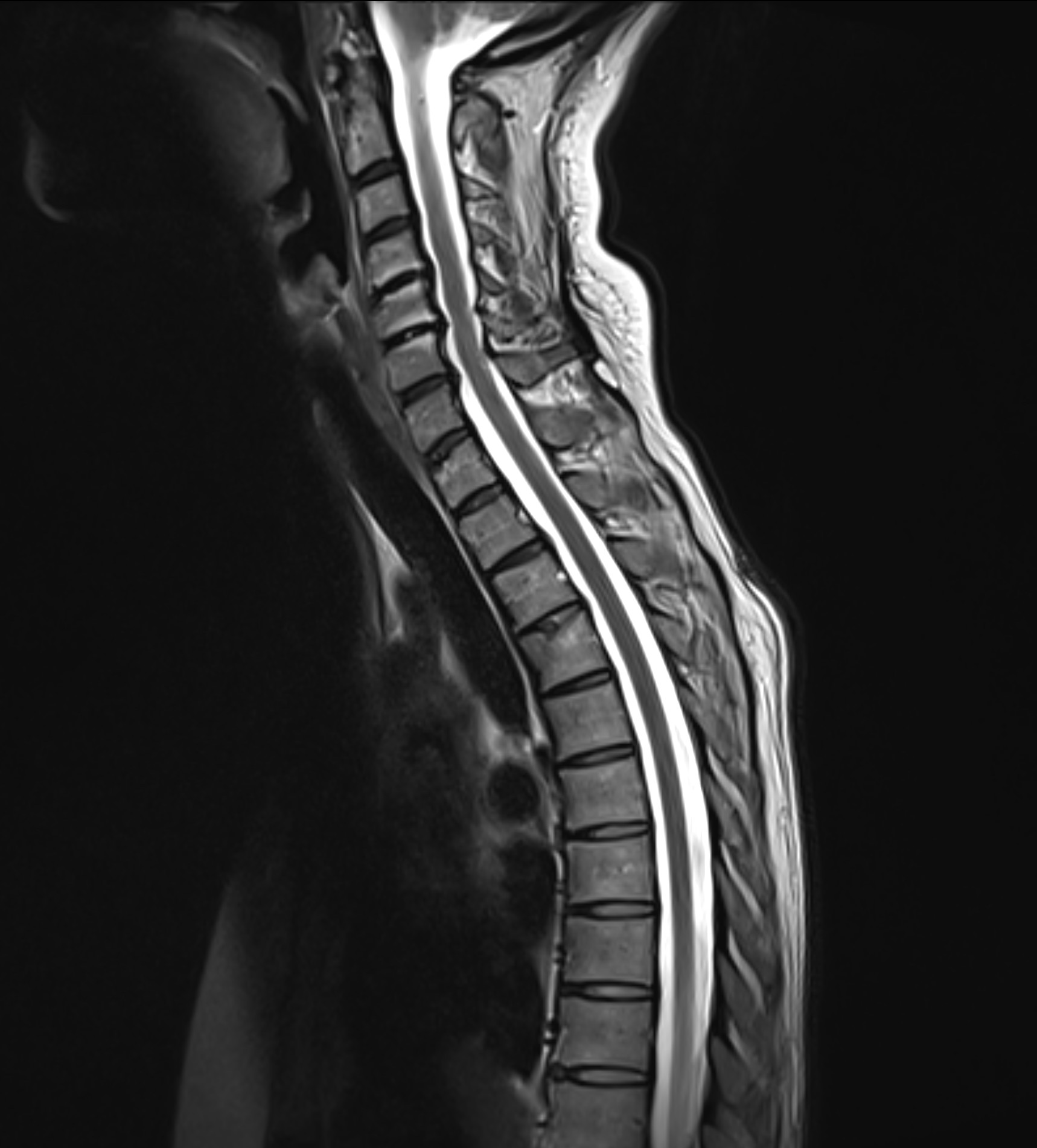
MRI of the entire spine – cervical, thoracic and lumbar
MRI Full Back is an advanced magnetic resonance imaging (MRI) examination that covers the entire spine – from the cervical spine and thoracic spine to the lumbar spine. The examination provides a detailed image of all parts of the spine and surrounding structures, making it possible to identify both acute and chronic changes. Since the examination covers all spinal segments, it is especially recommended for those who have more extensive or diffuse problems that cannot be localized to a specific part of the back.
The MRI examination is completely radiation-free, painless and provides high-resolution images with millimeter precision. The referral is sent immediately and you will receive a clear written opinion from an experienced radiologist based on the results.
When is an MRI of the back appropriate?
MRI of the whole back is particularly appropriate if you:
- Have long-term or recurring back problems that are not localized to a specific part (e.g. both in the neck and lumbar spine).
- Have symptoms that radiate into the arms or legs, numbness, weakness or affect nerve roots.
- Have complex problems that may be due to more than one spinal segment (e.g. suspicion of both herniated discs and spinal stenosis).
- Need to exclude or follow up more serious changes such as tumors, metastases or bone destruction.
- Have diagnosed diseases that can affect several parts of the back, such as Bechterew's disease (ankylosing spondylitis) or multiple sclerosis.
If you have clear problems only in a limited area, such as the lumbar or cervical spine, a more focused MRI examination of the specific area may be better suited.
What diseases and changes can be detected with a full back MRI?
A full back MRI can identify a wide range of diseases and changes in the spine. The examination provides a detailed picture of all spinal segments and can, among other things, demonstrate:
- Disc changes: Herniated discs, bulging discs and degenerative changes.
- Narrowings: Spinal stenosis and other congestion around the spinal cord and nerve roots.
- Joint diseases: Osteoarthritis and inflammatory changes in the facet joints.
- Skeletal changes: Scoliosis, skeletal destruction and spinal sclerosis.
- Nerve effects: Signs of pressure or irritation of nerve roots, for example in sciatica.
- Tumors and metastases: Primary tumors, metastases and other tissue changes.
- Inflammatory and neurological changes: Multiple sclerosis (MS plaques), Bechterew's disease and other inflammatory processes.
Important things to know before your MRI examination of the spine
Do you have a pacemaker, insulin pump or other electronic equipment in your body? It is important that you inform us before the examination. You do this by answering the questions in the health declaration you receive when you activate the referral in our test response service. Most metal implants, such as hip or knee replacements, are usually not an obstacle to performing an MRI examination of the spine, but we always need to receive information about these in advance.
After the examination
- You will receive a personal report in which an experienced radiologist and specialist doctor analyzes your MRI images and explains their meaning.
- You will receive a digital login with access to your X-ray images and report.
- If the examination shows serious changes, you will quickly receive a referral within healthcare for further investigation and treatment.
For those who want answers to their back problems
MRI Full Back is specially designed for those who have recurring or more extensive back problems and want a clear explanation of the cause. The examination can reveal early changes in the spine that are otherwise difficult to detect, and is completely risk-free and without harmful radiation.
Order and activate your referral immediately – you will usually receive a summons within 7–10 working days.


