What do you see on an MRI scan?
An MRI (magnetic resonance imaging) scan produces very detailed images of the body's internal structures – especially soft tissues, nerves, organs, joints and blood vessels. Strong magnetic fields and radio waves create cross-sectional images that enable doctors to detect injuries, diseases or changes that are not as clearly visible on regular X-rays or computed tomography (CT).
The doctor looks for signs of diseases and injuries in the area in question on the images. Tumors, circulatory disorders, inflammatory conditions, nerve compressions and other changes in the brain, spine, abdomen and musculoskeletal system can be detected.
MRI is particularly effective for examining:
- The brain: if a stroke, tumor, dementia, inflammation or multiple sclerosis is suspected.
- The spine: e.g. herniated disc, nerve damage or spinal stenosis.
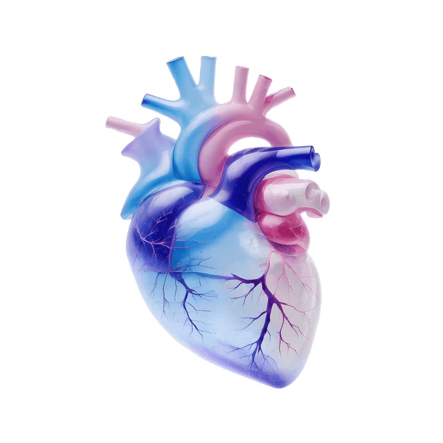
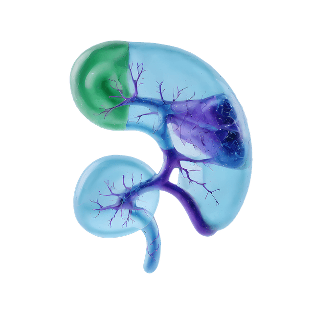
- Internal organs: such as liver, kidneys, pancreas and prostate – e.g. cysts, tumors or inflammation.
- Joints and skeleton: e.g. meniscus injuries, ligament injuries, osteoarthritis or stress fractures.
- Nerves: pinching, tumors or other changes in the nerve pathways.
Since MRI does not use X-ray radiation, but is based on magnetic resonance, the examination can be used for repeated imaging and provides exceptionally good contrast between different tissue types. This makes it possible to detect even very small changes in the body.

