Quick version
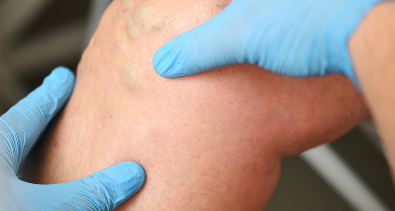
Knee injuries are common and can occur both during acute stress and as part of slow, degenerative changes. Symptoms such as pain, swelling, instability or reduced mobility can have several different underlying causes, with meniscus injuries, cruciate ligament injuries, cartilage damage and osteoarthritis being some of the most common.
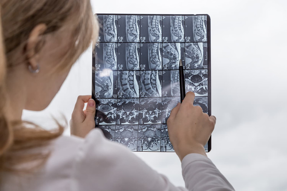
To ensure the correct diagnosis, it is important to have a thorough medical assessment. In the event of unclear or persistent complaints, an MRI examination can provide a detailed picture of the knee joint's structures and identify injuries that are not visible with traditional X-rays.
At Testmottningen you will receive a complete chain of care - from initial medical assessment and referral for MRI, to a statement from a specialist and follow-up review. This gives you a safe and clear path forward, regardless of whether it involves further treatment or your own rehabilitation.
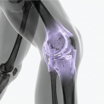
The knee joint is one of the body's most stressed joints and is exposed to great forces every day. This also makes it particularly vulnerable to both acute injuries and slow, degenerative changes. Pain in the knee can occur suddenly in connection with sports or accidents, but can also develop over time without a clear injury.
With a forensic investigation, it is often possible to identify the cause of the problem and initiate the right treatment in time. In many cases, magnetic resonance imaging (MRI) plays a central role in getting a clear picture of what is happening inside the knee.
Here are the 5 most common knee injuries
1. Meniscus injury
The meniscus acts as a shock absorber in the knee and is often damaged by twisting or straining. The injury can be acute, but is also common as part of age-related changes.
- Pain when straining or twisting.
- Locking or clicking sounds in the knee.
- Swelling after activity.
2. Cruciate ligament injury (ACL/PCL)
The cruciate ligaments stabilize the knee. The most common injury is the anterior cruciate ligament (ACL), often in connection with sports.
- Sudden pain and instability.
- Feeling that the knee is "giving way".
- Rapid swelling.
3. Ligament injuries (ligaments)
Injuries to the inner or outer ligaments often occur during trauma, such as when tackling or falling.
- Pain on the inside or outside of the knee.
- Swelling and tenderness.
- Reduced stability.
4. Cartilage damage
The cartilage in the knee joint can be damaged by strain or wear. This can lead to pain and stiffness.
- Pain when straining.
- Stiffness and reduced mobility.
- In some cases, swelling.
5. Osteoarthritis (degenerative changes)
Osteoarthritis involves the breakdown of joint cartilage and is common with increasing age. The changes can also include meniscus damage and inflammation in the joint.
- Nagging pain
- Stiffness, especially after rest
- Impaired function over time
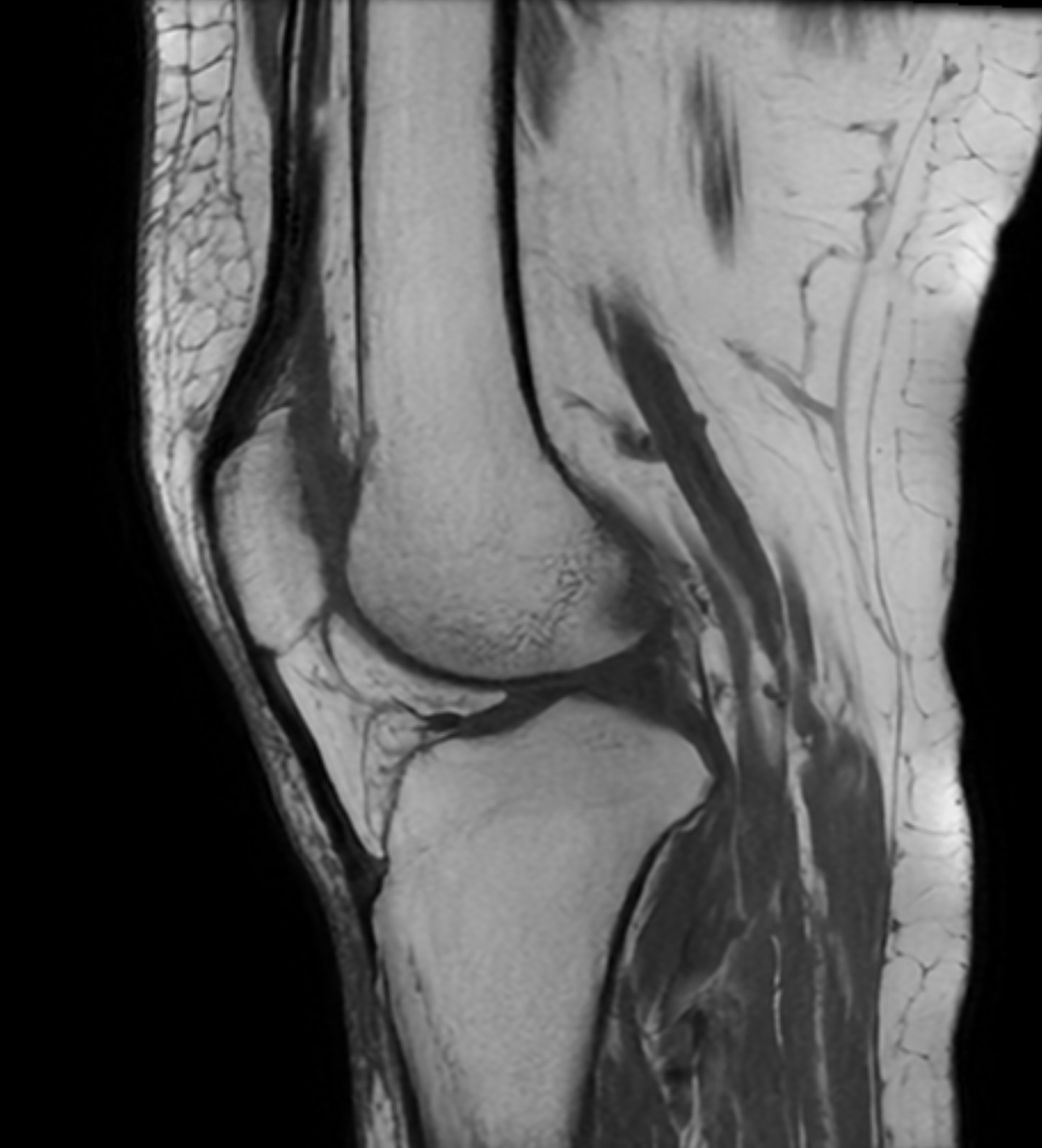
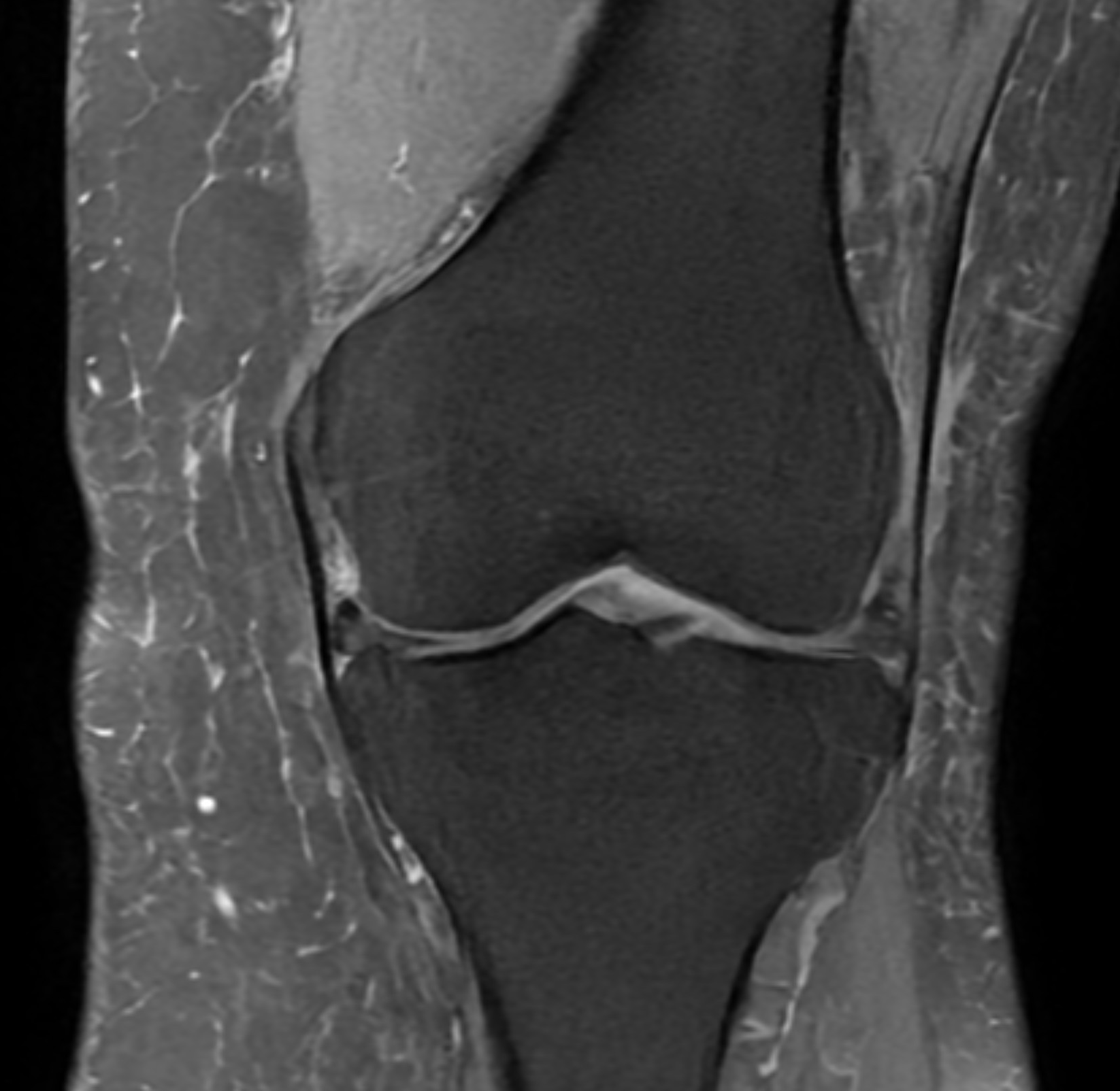
MRI of the knee – how it works
Magnetic resonance imaging (MRI) is an advanced imaging method that provides detailed images of the soft tissues of the knee joint, such as the menisci, cruciate ligaments, cartilage and muscles. Unlike X-rays, MRI can show damage that is not visible on traditional images.
The examination is completely radiation-free and is often used when a more accurate diagnosis is needed.
When MRI examination of the knee is relevant
Magnetic resonance imaging (MRI) of the knee is recommended when the clinical picture is unclear or when symptoms persist despite relief and initial treatment, such as rest or physiotherapy. MRI is particularly valuable for identifying injuries to the soft tissues of the knee joint, which cannot be visualized with conventional X-rays.
The examination is indicated in cases of suspicion of:
- Meniscal injuries, both traumatic and degenerative
- Cruculent ligament injuries (anterior or posterior cruciate ligament)
- Cartilage injuries or early signs of articular cartilage degeneration
- Inflammatory changes or increased amount of joint fluid (joint effusion)
MRI is also used as a basis for further treatment, such as planning physiotherapy, injection treatment or surgical procedures, and to assess the extent and prognosis of the injury.
Examples of MRI findings
An MRI examination can sometimes show several changes in the knee at the same time. An example is degenerative meniscus injuries, where the tissue has changed over time and may have cracks (ruptures).
In some cases, the following is also seen:
- Preserved or slightly affected articular cartilage
- Discrete changes in the cruciate ligament without total rupture
- Increased amount of synovial fluid (signs of irritation)
- Smaller cysts, such as Baker's cyst
These always need to be interpreted in relation to symptoms to determine the significance and possible treatment.
This is how it works with us
At Testförmedlingen, we ensure that you receive the right investigation from start to finish.
- Assessment by a general practitioner before a referral is sent
- MRI examination at an affiliated clinic
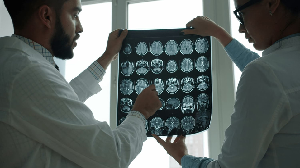
- Report from a specialist (radiologist)
- Follow-up and review with a general practitioner
This means that you will not only receive images – but a clear medical interpretation and guidance on the next steps.
When should you seek help?
You should consider further investigation if you have:
- Pain that does not go away
- Recurrent swelling
- Instability in the knee
- Difficulty putting weight on or moving the knee normally
Early diagnosis can make a big difference ideal for both treatment and recovery.