Quick version
Understanding what is happening in the body without cutting or radiation is one of the great benefits of modern medicine. With an MRI – or magnetic resonance imaging – the doctor gets a detailed look at the body's inner world, from the convolutions of the brain to the abdominal organs and the spinal nerve pathways. The technology is used every day in healthcare to detect everything from inflammation and tumors to circulatory disorders and nerve damage.
What makes MRI so unique is that it provides high-resolution images of both soft tissues and tissue – completely without radiation. That's why it is often used in investigations of problems where other methods are not sufficient. This can involve understanding why someone has a headache or dizziness, why back pain won't go away, or what is really going on in a joint that won't heal.
What is an MRI scanner?
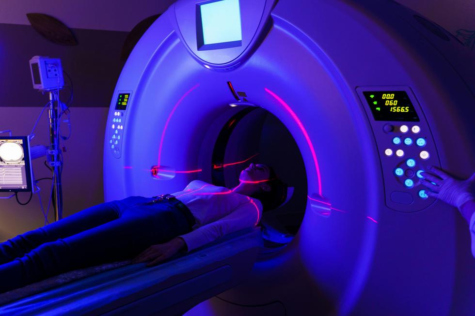
A MRI scanner, or MRI scanner, is an advanced medical device used to create highly detailed images of the inside of the body – without using X-rays. The technology is based on powerful magnetic fields and radio waves, which affect the body's hydrogen atoms and convert the signals into high-resolution images.
Because MRI primarily visualizes soft tissues, organs, vessels and nerves, it is one of the most valuable methods for investigating conditions where regular X-rays or ultrasound are not sufficient. The images are reviewed by specialized doctors who can see even very small changes in, for example, the brain, spine, joints or abdomen. MRI is a safe and completely painless examination that can be performed both in the event of acute symptoms and as part of long-term follow-up.
Below we will review some of the most common MRI examinations and what they are used for and what they can identify.
The brain
MRI of the brain is one of the most common examinations that doctors order, especially for symptoms such as recurring headaches, dizziness, visual disturbances, numbness or memory problems. The examination provides very detailed images of the brain's tissues, vessels and nerve pathways, which makes it possible to detect even small changes that can be of great importance for the diagnosis.

With an MRI camera, you can identify tumors, cysts, inflammation (such as in MS), signs of stroke, bleeding and increased pressure in the brain. It is also an important tool when suspecting neurodegenerative diseases such as Parkinson's or Alzheimer's.
MR brain is used both in acute conditions and to follow up on previously known changes. The examination is radiation-free, completely painless and is considered one of the most accurate methods for assessing the function and structure of the brain.
Lower back
The lower back is a particularly vulnerable part of the spine, as it supports the weight of the upper body and is stressed during everything from lifting to sitting. Pain in the area can be due to several different causes – from wear and tear in the discs to pressure on the nerves that cause sciatica. An MRI for lower back allows you to study the discs, nerve roots, joints and spinal cord in detail, which is crucial when regular X-rays are not enough. The examination can explain why you have pain that radiates down your leg, why your sensation has changed or why your mobility is limited. It also makes it possible to detect inflammation, infections or changes after previous back problems.
Knee
The knee and knee joint are one of the body's most complex and injured joints – especially in people who are physically active. When pain, swelling or instability occurs, it is often difficult to know exactly which structure is affected. An MRI examination of the knee makes it possible to map injuries to the cruciate ligaments, meniscus, ligaments, cartilage and joint capsule with high precision.
In many cases, it is an invaluable tool in cases of suspected sports injuries or when you want to rule out inflammation, cartilage breakdown or early osteoarthritis. The examination is also important before possible surgery or as a basis for rehabilitation planning.
Prostate
The prostate gland can change with age, but certain changes can also be signs of more serious conditions. In the case of elevated PSA values, difficulty urinating or suspected cancer, a
magnetic resonance imaging of the prostate
is useful for mapping tissue changes in the different zones of the prostate. The examination can identify suspected tumor areas that are otherwise difficult to detect, and often helps reduce the need for random biopsies. It is used both in primary investigation and for follow-up of previous findings, as well as as decision support for treatment.How to book an MRI
You start by choosing the type of MRI examination you want performed. When your order is completed, we automatically send a referral to one of our affiliated X-ray units based on any symptoms and a completed health declaration.
Within a few working days, you will receive a summons with the time and place for the examination. The MRI examination itself is performed by licensed personnel and is painless and free of radiation. Afterwards, the images are carefully reviewed by a specialist doctor, and you will receive a written report within 2 to 5 days. The examination takes between 30-50 minutes.
You don't have to go through a health center or stand in line – the entire process takes place on your terms, with a focus on medical quality and accessibility.


