


MRI Knee – Detailed assessment of the knee joint for pain, swelling, or suspected injury
The knee joint is the largest and most heavily loaded joint in the body – a complex structure made up of bones, cartilage, ligaments, menisci, tendons, and bursae that together enable movement, stability, and shock absorption. This makes the knee particularly vulnerable to both acute injuries and wear-and-tear.
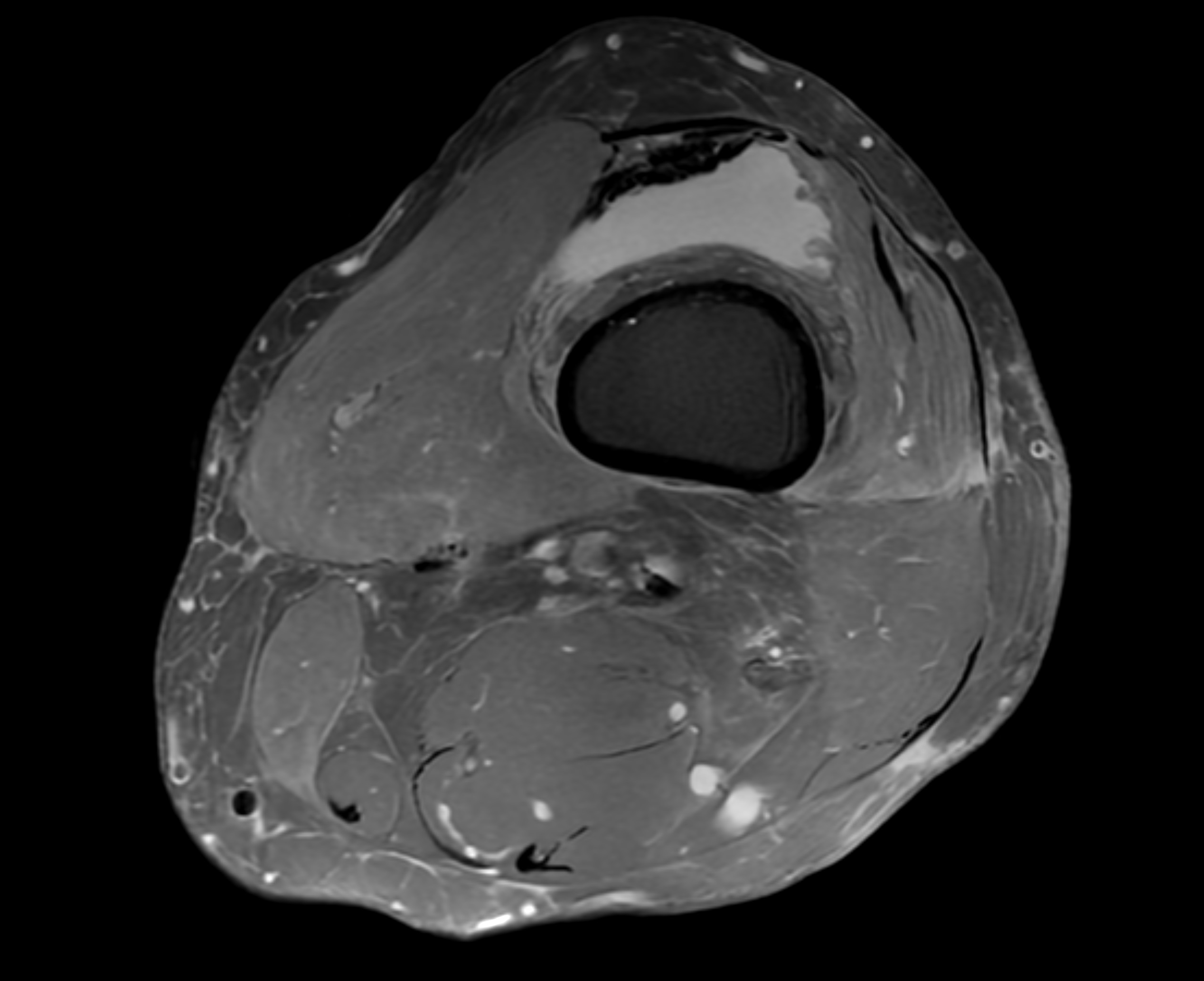
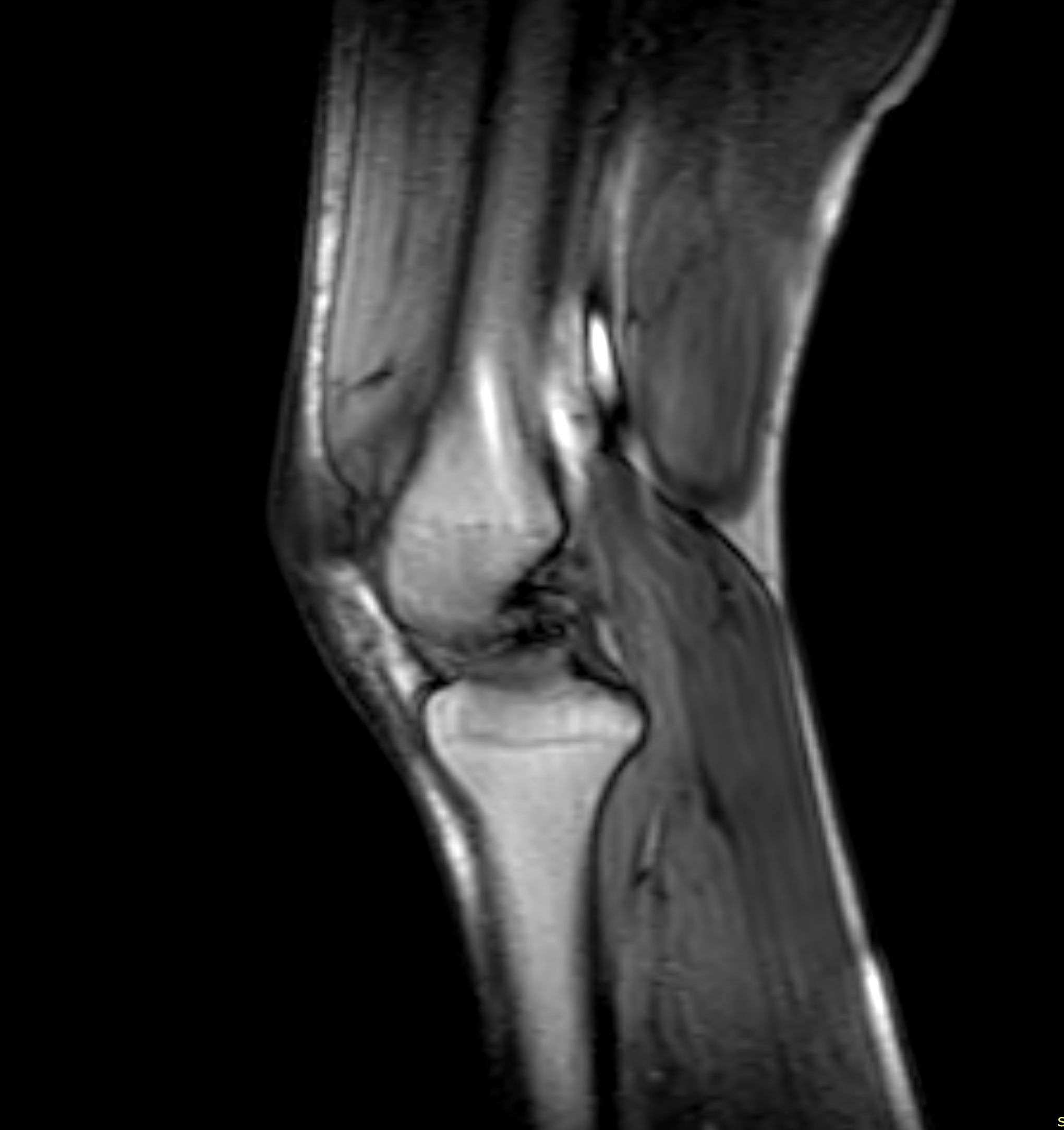
An MRI (magnetic resonance imaging) of the knee or knee joint provides highly detailed images of both bone and soft tissue. The examination is radiation-free, painless, and superior to other methods for detecting meniscal tears, ligament injuries, cartilage damage, arthritis, or tendon inflammation. MRI of the knee is especially recommended for persistent pain, swelling, instability, or if you suspect an internal knee injury following sports activity, twisting, or trauma. You can book directly – no referral needed – and receive an appointment within a few days.
When is an MRI of the knee recommended?
Knee pain can result from a wide variety of causes, including trauma, overuse, degenerative changes, or inflammation. In some cases, clinical examination, X-ray, or ultrasound may not be sufficient to detect damage to the meniscus, ligaments, or cartilage – and MRI becomes the preferred alternative.
You should consider an MRI of the knee if you experience:
- Pain during load or movement – especially during bending, stair climbing, or physical activity.
- Swelling, warmth, or stiffness in the knee – may indicate joint inflammation, bursitis, or fluid accumulation.
- Instability or "giving way" of the knee – often seen with ligament or cruciate injuries.
- Locking or clicking sensations – possible signs of meniscal injury or cartilage fragments.
- Pain following a sports injury, fall, or twisting motion – particularly if rest or physiotherapy has not led to improvement.
MRI of the knee is particularly valuable in several clinical situations. If you are preparing for surgery – for example, due to a suspected meniscus or cruciate ligament injury – MRI provides a precise map of the internal structures of the knee, which is critical for surgical planning.
The examination is also important when previous imaging, such as plain X-rays or ultrasound, has been inconclusive. This is especially true for soft tissue injuries, cartilage damage, or difficult-to-assess pain where an internal lesion is suspected.
MRI is also used to reach an exact diagnosis when symptoms do not respond to treatment or when more information is needed to determine the next step – whether it's physiotherapy, injection, or surgery. In such cases, MRI provides a safe and accurate path to the right care.
MRI is commonly used to evaluate the following knee conditions
- Meniscal tear – Injury to the medial or lateral meniscus can cause locking, pain, or swelling in the knee. MRI helps determine whether it is a small fissure, a full tear, or age-related degeneration.
- Anterior or posterior cruciate ligament injury (ACL/PCL) – Common in sports-related injuries. MRI is the best method for visualizing whether the ligament is stretched, partially torn, or completely ruptured.
- Collateral ligament injuries (MCL/LCL) – These ligaments stabilize the knee from the sides. MRI detects sprains, tears, or ruptures and can also reveal associated injuries.
- Cartilage damage and osteoarthritis – MRI detects early signs of cartilage thinning, irregularities, cracks, and early osteoarthritis that are not visible on X-ray.
- Bursitis – Often located behind the kneecap or on the inside of the knee. MRI shows fluid accumulation and inflammatory changes in the bursae.
- Bone marrow edema and stress fractures – Can occur from overuse, especially in athletes. MRI detects fluid in the bone or microfractures before a full fracture develops.
- Rheumatoid arthritis and synovitis – Inflammation in the joint capsule or early bone erosion can be seen clearly on MRI, often long before changes appear on X-ray. This is important in rheumatological evaluation.
- Plica syndrome – Inflammation of folds in the joint lining that may cause pain, snapping, or motion restriction. MRI confirms the diagnosis and helps rule out other causes.


