MR Aorta – MRI of the body's largest vessel
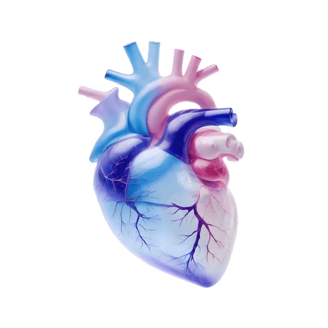
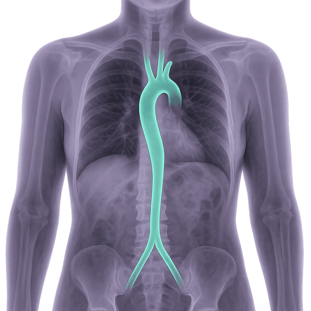
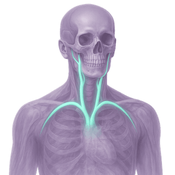
MR Aorta including Vena cava is an MRI angiography that maps the body's largest artery and veins. The aorta transports oxygenated blood from the heart out into the body, while the vena cava returns oxygen-poor blood to the heart. Changes in these vessels can lead to serious conditions and often need to be detected in time to prevent complications.
The examination provides high-resolution sequences of the entire aorta and the superior and inferior vena cava. With the information, the radiologist can identify enlargements (aneurysms), tears in the vessel wall (dissection), narrowings, blockages or external pressure that affects blood flow. The examination is radiation-free, painless and a very accurate diagnostic tool for suspected vascular disease.
When is MRI of the aorta and vena cava recommended?
An MRI is recommended when there is suspicion of vascular diseases that may affect circulation or cause serious symptoms. It is also used to follow up on known changes or before surgical procedures.
Common symptoms and complaints that may justify the examination:
- Chest or back pain with suspected vascular origin.
- Throbbing sensation or swelling in the abdomen.
- Unexplained shortness of breath, swelling of the face, neck or arms.
- Signs of reduced blood flow or suspected blockage.
- Follow-up of previously detected aortic aneurysm or dissection.
- Investigation in case of elevated cholesterol levels or other cardiovascular risk profile.
Conditions when MRI of the Aorta and Vena Cava is recommended
- Aortic aneurysm dilation of the aorta that may be at risk of rupture.
- Aortic dissection tear in the vessel wall that can be life-threatening.
- Atherosclerotic changes narrowing and hardening of the arteries that affect blood flow.
- Vena cava syndrome compression or blockage of the vena cava that can cause swelling, respiratory distress or discomfort.
- Thrombosis blood clots in the large veins.
- Tumors or metastases that press on or grow into the aorta or vena cava.
- Malformations or congenital vascular anomalies that affect circulation.
Book MRI Aorta and Vena cava – get a referral immediately
An MRI examination of the aorta and vena cava is a central diagnostic tool for detecting and mapping vascular diseases such as aneurysms, dissections, thromboses or other abnormalities that can affect blood flow. The examination provides detailed cross-sectional images of the vascular wall and surrounding structures, which enables a careful assessment of the extent and severity of the disease. MRI is particularly valuable in situations where you want to avoid ionizing radiation or where other imaging diagnostics have not provided sufficient information. The results are reviewed by a specialist in radiology who compiles a written report that can form the basis for further medical management and treatment decisions.


