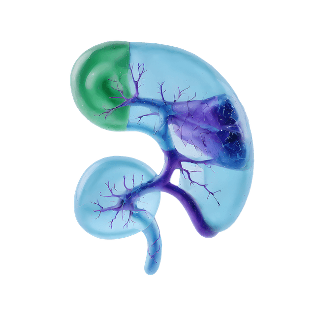
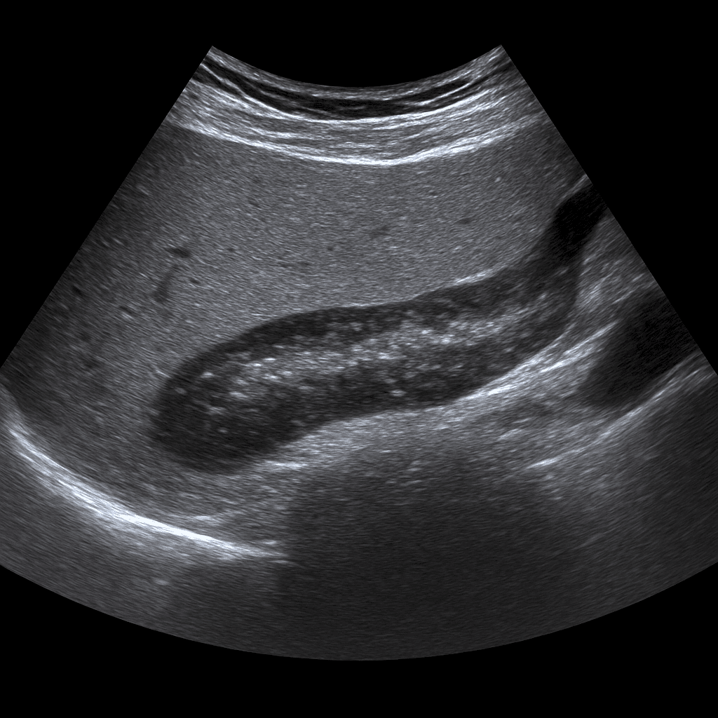
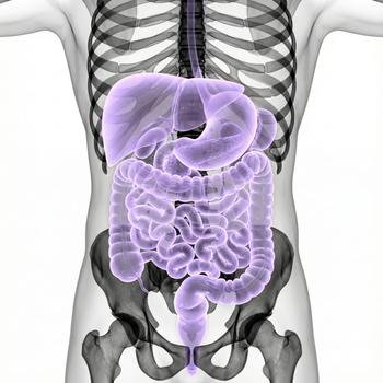
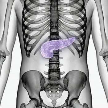
An ultrasound examination of the pancreas or (pancreas) is used to examine the size, shape, tissue structure and surrounding organs of the gland. The examination is performed by a specialist in radiology and provides real-time images that can show changes caused by inflammation, cysts, stones or tumors. Ultrasound of the pancreas is often used as the first step in the investigation of abdominal pain, jaundice or abnormal liver and pancreatic values in blood tests.
Ultrasound of the pancreas - for pain, jaundice or elevated enzyme values
The examination is recommended for diffuse or recurrent abdominal pain, nausea, unexplained weight loss, jaundice or elevated levels of amylase, lipase or liver tests. It is also used to follow up on previous inflammation (pancreatitis) or cystic changes in the pancreas.
Unlike MRI and CT, which are used for more detailed mapping of glandular tissue and the urinary system, ultrasound is a fast and radiation-free method for detecting major structural changes, fluid accumulation, and surrounding inflammation. Ultrasound can sometimes be limited in the case of gas-filled intestines or deep abdominal tissue, but works excellently as an initial examination and for follow-up of known findings.
Common symptoms and questions
- Pain in the upper abdomen or radiating to the back.
- Jaundice or itching of the skin (signs of impaired bile or pancreatic outflow).
- Elevated levels of amylase, lipase or liver tests.
- Suspected pancreatitis (inflammation of the pancreas).
- Unexplained weight loss or loss of appetite.
- Follow-up of previous cysts or changes in the pancreas.
Conditions that can be detected with ultrasound of the pancreas
- Pancreatitis – acute or chronic inflammation of the pancreas.
- Pancreatic cysts or pseudocysts.
- Changes in the gait system due to obstruction or swelling.
- Fat deposits or calcifications in chronic inflammation.
- Tumor or suspicious mass that requires further investigation with MRI or CT.
- Fluid accumulation or reactive changes in surrounding tissue.
How an ultrasound of the pancreas is performed
The examination is performed while you lie on your back or left side. A gel is applied to the skin and the doctor moves the ultrasound probe over the upper part of the abdomen, usually under the left rib cage. The examination usually takes 15–20 minutes. For the best image quality, it is recommended that you fast for about 4–6 hours before the examination to reduce the amount of air in the stomach and intestines.
Order an ultrasound examination of the pancreas – get a statement and recommendation from a doctor
The images are reviewed by a specialist in radiology who draws up a written medical report. The answer is delivered digitally within a few working days and can be shared with your treating doctor for further investigation or treatment. If the ultrasound shows unclear or deeper changes, it is often recommended to supplement with MRI or CT for more detailed mapping.

