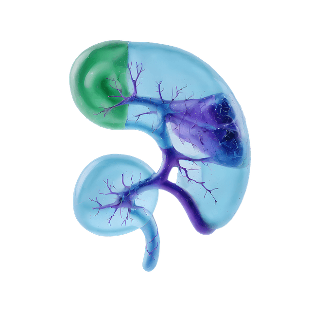
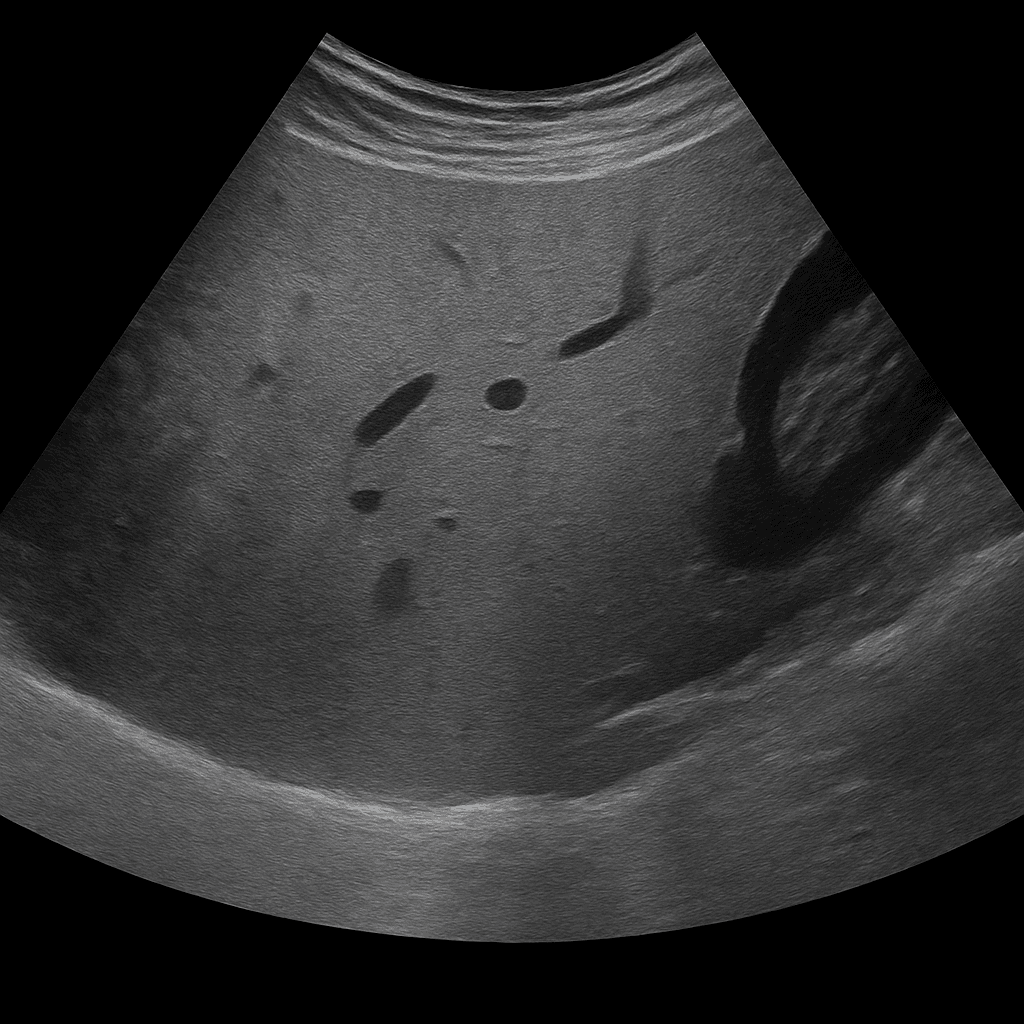
Ultrasound examination of the liver is used to examine the size, structure and vascular flow of the liver. The examination is performed by specialist radiologists and provides detailed images in real time that show changes that may indicate fatty liver, inflammation, cysts, tumors or cirrhosis. Ultrasound of the liver is often the first-line method for investigating abnormal liver tests or abdominal complaints related to the liver.
Ultrasound of the liver - for abdominal pain, elevated liver values or suspected fatty liver
Ultrasound of the liver is recommended for elevated liver values in blood tests (e.g. ALT, AST, ALP or GT), abdominal pain, fatigue or if fatty liver (steatosis) is suspected. The examination is also used to follow up on known liver diseases such as hepatitis, cirrhosis or previous findings of cysts and tumors.
Unlike MRI or CT, which are used for detailed mapping of tissue changes and blood vessels, ultrasound is the primary method for assessing liver structure, fat deposition, vascular flow, and volume. Ultrasound can quickly detect abnormalities without radiation and is often used as the first step in liver diagnostics.
Common symptoms and questions
- Elevated liver values in blood tests (ALT, AST, ALP, GT).
- Suspected fatty liver (steatosis) or liver inflammation (hepatitis).
- Pain or pressure under the right rib cage.
- Fatigue, nausea or unexplained weight loss.
- Follow-up of known cysts, tumors or cirrhosis.
- Check for alcohol exposure, drug treatment or infection.
Conditions that can be detected with liver ultrasound
- Fatty liver (steatosis) – increased accumulation of fat in the liver tissue.
- Liver enlargement (hepatomegaly) due to inflammation or cirrhosis.
- Cysts, hemangiomas or other benign changes.
- Signs of fibrosis or cirrhosis (uneven surface and altered structure).
- Tumor or suspected metastasis in the liver.
- Impaired blood circulation in the hepatic veins or portal veins (portal hypertension).
How an ultrasound of the liver is performed
The examination is performed while you lie on your back or slightly on your left side. A gel is applied to the skin and the doctor moves the ultrasound probe over the area under the right rib cage. The examination usually takes 15–20 minutes. For the best image quality, you should fast for about 4–6 hours beforehand, as food and air in the intestine can affect the clarity of the image.
Order an ultrasound examination of the liver - get a doctor's opinion and recommendation
The images are reviewed by a specialist in radiology who draws up a written medical opinion. The answer is delivered digitally within a few working days and can be shared with your treating physician for continued follow-up or treatment. If necessary, the examination can be supplemented with MRI or CT for more detailed mapping of liver tissue and vessels.

