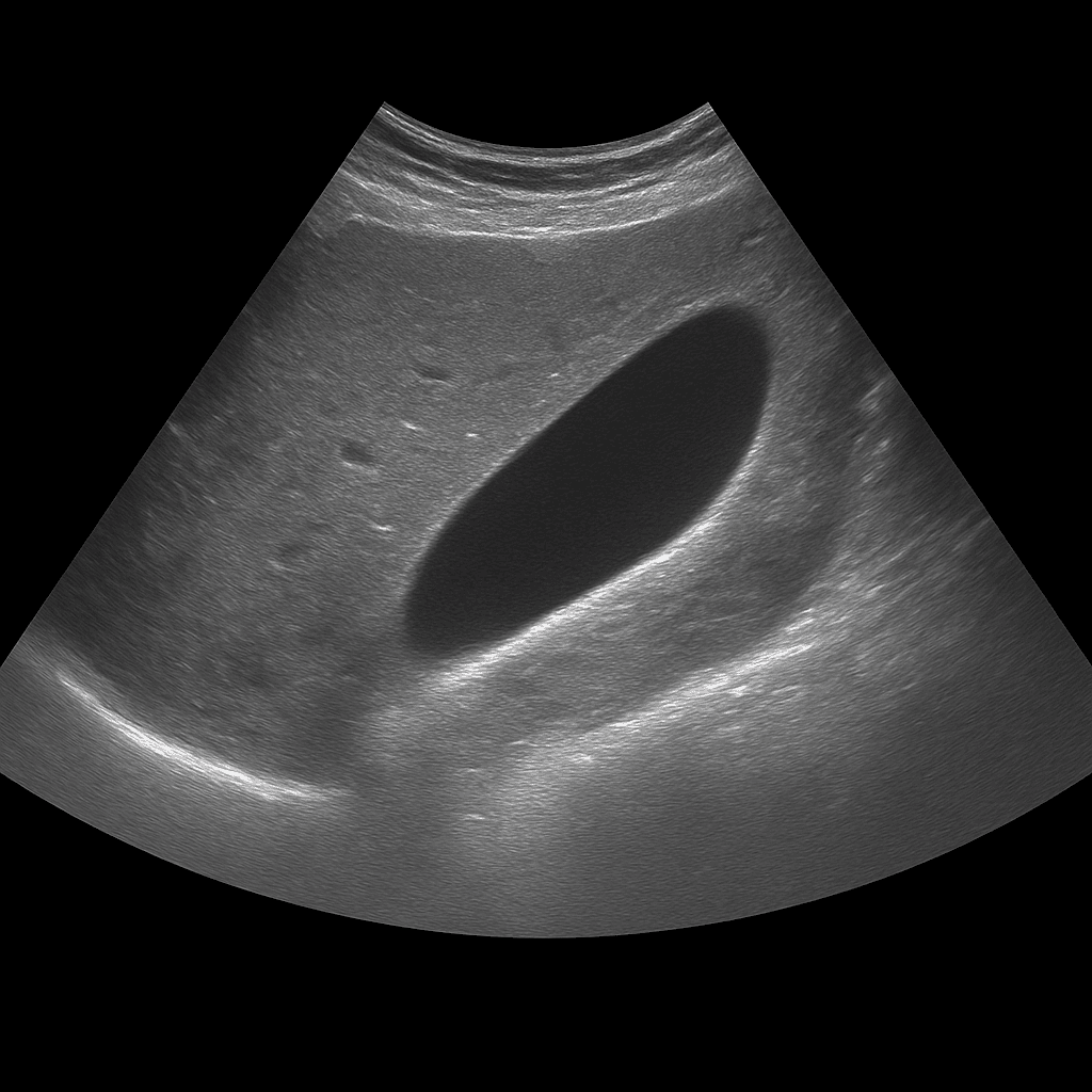
A gallbladder ultrasound is used to examine the gallbladder wall, contents and function. The examination is performed by a specialist in radiology and provides detailed real-time images that show whether the gallbladder contains stones, fluid or signs of inflammation. Gallbladder ultrasound is used as the first-line method when gallstone disease or inflammation of the gallbladder (cholecystitis) is suspected.
Gallbladder ultrasound – for pain, nausea or suspected gallstones
A gallbladder ultrasound is recommended for pain under the right rib cage, nausea, bloating or recurrent discomfort after eating fatty foods. The examination can show whether the gallbladder contains stones, sludge (biliary gravel) or a thickened wall that indicates inflammation. It is also used to follow up on previous gallstone problems or after surgery where parts of the biliary system remain.
Unlike biliary tract ultrasound, which focuses on the bile ducts and their connection to the liver, gallbladder ultrasound focuses on the wall, contents, and function of the gallbladder itself. The method is used to detect acute or chronic changes in the organ, while MRI or CT is used when the bile ducts and surrounding organs need to be assessed in more detail.
Common symptoms and questions
- Pain under the right rib cage, often after meals.
- Nausea, bloating or vomiting when eating fat.
- Recurrent gallstone attacks or previous gallstone disease.
- Suspected acute or chronic cholecystitis (gallbladder inflammation).
- Unclear abdominal complaints with suspicion of gallbladder changes.
- Follow-up after previous surgery or gallbladder removal.
Conditions that can be detected with gallbladder ultrasound
- Gallstones or sludge (biliary gravel) in the gallbladder.
- Acute or chronic cholecystitis – inflammation of the gallbladder wall.
- Thickened gallbladder wall or fluid around gallbladder.
- Polyps or small mucosal changes in the gallbladder.
- Gallbladder hydrops – distended gallbladder due to obstruction in the descending duct.
- Cysts, tumors or other focal changes in the gallbladder.
How an ultrasound of the gallbladder is performed
The examination is performed while you lie on your back or slightly on your left side. A gel is applied to the skin and the doctor moves the ultrasound probe over the area under the right rib cage. The examination usually takes 10–20 minutes. For the best image quality, you need to fast for at least 4–6 hours beforehand, as an empty gallbladder is difficult to assess.
Order an ultrasound examination of the gallbladder – get a doctor's opinion and recommendation
The images are reviewed by a specialist in radiology who draws up a written medical opinion. The answer is delivered digitally within a few business days and can be shared with your treating physician for further investigation or treatment. If necessary, the findings can be followed up with MRI or CT for more detailed assessment of the biliary tract or liver.

