Quick version
The knee joint is a vital structure for movement and stability and is often subject to injury and wear and tear during sports or overuse.
- Connects the femur, tibia, and kneecap
- Stabilized by ligaments, menisci, and muscles
- Common site of sports-related injuries
- Arthritis and inflammation can cause chronic pain
- Diagnosis includes clinical tests and imaging techniques
What is the knee joint?
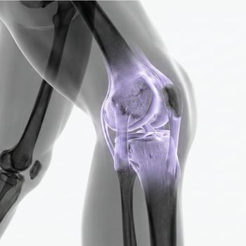
The knee joint (articulatio genus) is a complex joint that connects the thigh bone (femur) to the shin bone (tibia) and kneecap (patella). It allows for bending and extending the leg and is essential for walking, running and jumping. The knee joint is stabilized by several ligaments, menisci and muscles that together provide freedom of movement and protection against injury.
Anatomy of the knee joint
The knee joint consists of three parts: the femorotibial joint (femur–tibia), the femoropatellar joint (femur–patella), and a network of structures that include the cruciate ligaments (ACL and PCL), collateral ligaments, menisci and joint capsule. Together they create a stable but mobile structure.
Movement and function
The knee joint mainly allows bending (flexion) and stretching (extension), but also some rotation when the leg is bent. Muscles such as the quadriceps (front of the thigh) and hamstrings (back of the thigh) control these movements and protect the joint from overload.
Protective structures
The menisci act as shock absorbers and help distribute the load in the joint. The kneecap enhances the power transmission of the thigh muscles and protects the joint in the event of a fall. The ligaments provide stability and prevent abnormal movement.
Common conditions and diseases
Pain in the knee joint can be caused by meniscus injuries, ligament tears, cartilage damage, knee osteoarthritis or inflammation. Injuries often occur during sports or sudden twisting, while osteoarthritis develops gradually with age or obesity.
Examination and diagnosis
Knee problems are investigated with a clinical examination of mobility, swelling and stability. Imaging tests such as X-rays, MRI of the knee joint or ultrasound are used to visualize internal structures and make an accurate diagnosis.
Relevant symptoms
- Pain when walking, climbing stairs or carrying weight
- Swelling and stiffness in the knee
- Clicking or locking sounds when moving
- Instability or a feeling that the knee is "giving way"
- Limited mobility and difficulty extending the leg
Related conditions and diagnoses
- Anterior cruciate ligament injury (ACL injury)
- Meniscus injury
- Knee osteoarthritis
- Patellar luxation (dislocated kneecap)
- Runner's knee


