Quick version
Anatomy of the Sacroiliac Joints
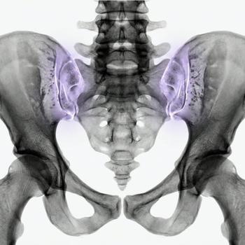
The sacroiliac (SI) joints are located in the lower part of the spine (os sacrum) and are strong, these joints connect the spine to the two hip bones (os ilium). These joints are crucial for supporting the weight of the upper body and transferring load to the legs when walking, standing and lifting.
Anatomical structure
The SI joints are partly synovial and partly fibrous, which makes them unique. By synovial we mean that they contain a cavity filled with synovial fluid (synovia), these joints are very mobile. Fibrous joints lack a joint cavity and synovial fluid, these joints are very stable but have limited or no mobility at all. The joints contain:
- Articular surfaces – covered by cartilage and shaped to limit movement but allow some sliding and shock absorption.
- Ligaments – very strong ligaments (including the anterior and posterior sacroiliac ligaments) stabilize the joint and prevent excessive movement.
- Close relationship to nerves – e.g. the sciatic nerve, which means that SI joint problems can cause radiating pain.
Common causes of SI joint pain
Pain from the SI joints can arise from many different causes, not just inflammatory ones. Some common conditions include:
- Mechanical dysfunction – e.g. oblique loading, leg length discrepancy, or pelvic ring instability.
- Overload – during prolonged standing, heavy work or incorrect posture.
- Pregnancy-related changes – hormonal softening of ligaments and increased mobility in the pelvis can cause pain.
- Post-traumatic conditions – after falls or accidents where the pelvis has been subjected to heavy load.
- Degenerative changes – cartilage loss and altered joint structure as part of natural aging.
How can problems and pain in the sacroiliac joints be investigated?
If it is suspected that the pain has an inflammatory origin or is part of a rheumatic disease, blood tests are an important part of the investigation:
- SR and CRP – general markers for inflammation in the body.
- HLA-B27 – genetic marker strongly linked to ankylosing spondylitis and other spondylarthritis.
- ANA and RF – to rule out other autoimmune diseases, such as SLE or rheumatoid arthritis.
- Liver- and kidney tests – may be needed for certain medication or as part of a comprehensive assessment.
Complementary imaging diagnostics such as an MRI of the sacroiliac joints can provide important information about early inflammatory changes, structural damage or other causes of pelvic and back pain that are not visible on regular X-rays.
In addition to MRI, other methods may sometimes be relevant depending on the symptoms and question:
- CT (computed tomography) – good for detecting fractures, bone spurs or previous fusions.
- Skeletal scintigraphy – sometimes used when widespread bone inflammation is suspected.
- Ultrasound – can be helpful in guiding injections or assessing nearby soft tissues.
A correct diagnosis is often based on a combination of clinical examination, imaging diagnostics and laboratory tests to understand whether the symptoms are mechanical, inflammatory or degenerative in nature.

