Quick version
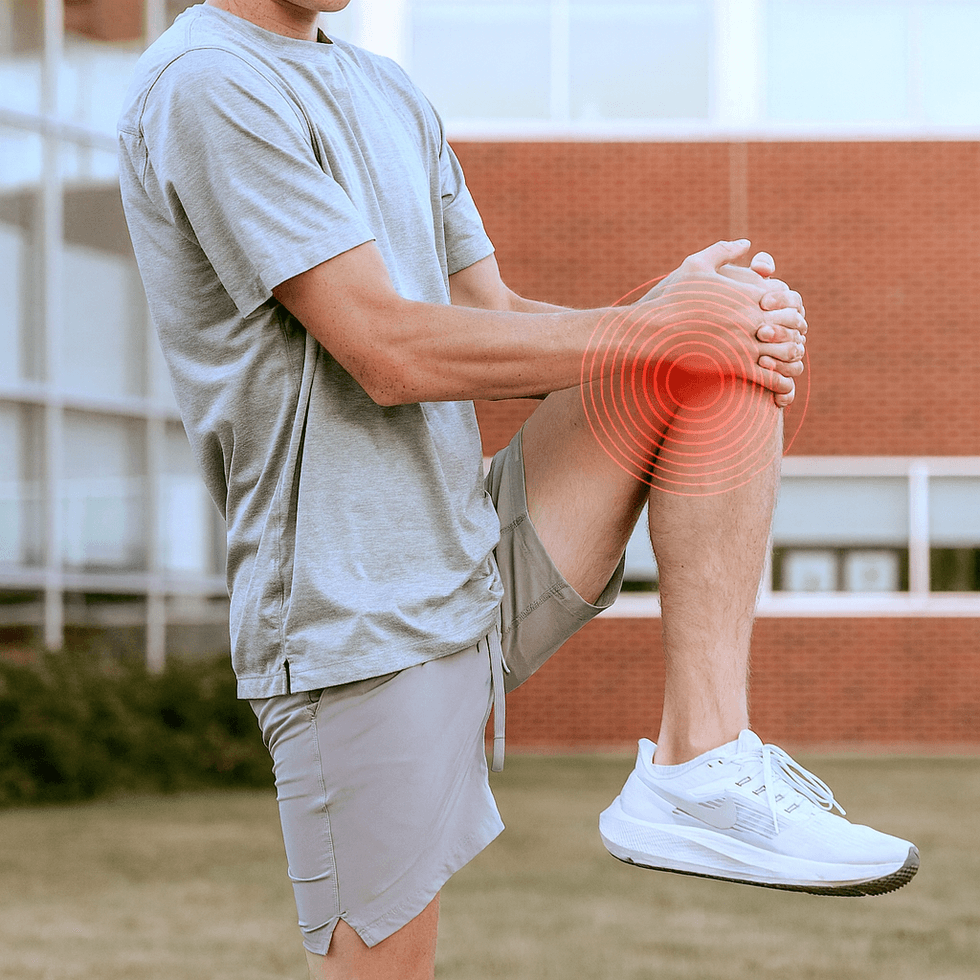
Knee pain is a very common condition that can affect all ages – from young athletes to older people with wear and tear problems. An MRI of the knee is the most accurate way to discover the cause of the pain, especially if meniscus damage, ligament damage or osteoarthritis is suspected.
If you want to get answers quickly and without a referral, you can book a private examination MRI Knee with us today.
In younger people, pain is often caused by overload, sports injuries or acute trauma, while older people are more often affected by wear and tear injuries (arthritis) and cartilage breakdown. Among active sportspeople, knee injuries account for almost 40% of all sports-related injuries, especially in football, handball, floorball, skiing and running.
Regardless of whether the pain comes on gradually or occurs after a torque, it is important to investigate the cause – and here an MRI of the knee plays a central role in identifying damage to the meniscus, cruciate ligaments, cartilage and other soft tissues in the joint.
But what actually happens in the knee when pain occurs
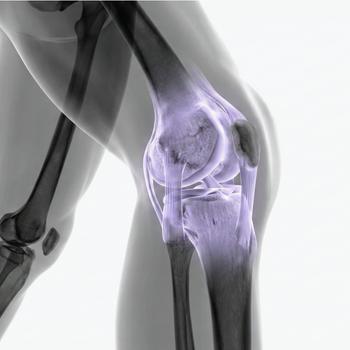
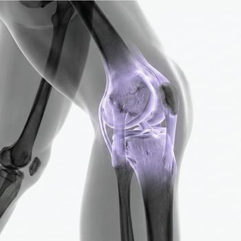
The knee joint is the body's largest and most complex joint, it supports almost the entire body weight when walking, jumping and running, and acts as a shock absorber with every step you take. The joint consists of three main bones: the thigh bone (femur), the shin bone (tibia), and the kneecap (patella). These are held together by a network of cartilage, ligaments, menisci, tendons, muscles, and bursae, which together allow the bone to bend, extend, and stabilize during movement.
Cartilage – the body’s shock-absorbing surface
The surface where the bones meet is covered by hyaline articular cartilage, a smooth and elastic layer of tissue that allows the bones to slide against each other without friction. When cartilage is worn or damaged, as in osteoarthritis, pain and stiffness occur as the bone surfaces begin to rub against each other.
The menisci – the joint’s balancer
Between the femur and tibia are two crescent-shaped cartilage discs, the medial and lateral menisci. They act as shock absorbers and stabilize the joint when stress is applied. A meniscus injury can cause sharp pain, swelling and a feeling that the knee is locking.
Ligaments and cruciate ligaments – stability and control
The knee joint is stabilized by four large ligaments: two collateral ligaments on the sides and two cruciate ligaments (anterior and posterior) in the middle of the joint. These prevent the knee from moving in the wrong direction. If a cruciate ligament ruptures – often as a result of a sudden torque – immediate pain, instability and swelling occur.
Tendons and muscles – the engine of movement
The large thigh muscles, mainly the quadriceps at the front and the hamstrings at the back, attach to the knee via strong tendons. The patellar tendon connects the kneecap to the shinbone. Inflammation or overload in these tendons can lead to pain at the front of the knee, often called jumper's knee.
Sacs and synovial fluid – the body's natural lubrication
Around the knee are several small sacs (bursae) that reduce friction between tendons, muscles and bones. When these are repeatedly stressed, they can become inflamed – a bursitis – which causes local swelling, redness and warmth.
When any of these structures is damaged, overloaded or inflamed, pain occurs as the body's warning signal. Knee pain can be sharp and sudden after a trauma, or diffuse and dull with long-term wear and tear. By understanding which tissue is affected, you can determine which treatment or examination – for example MRI examination of the knee – is needed to make the correct diagnosis.
Common causes of long-term knee pain
Overexertion and strain injuries
Prolonged strain or repetitive movements, such as running, jumping or cycling, can cause micro-damage to tendons and muscle attachments. Common diagnoses are runner's knee (iliotibial band syndrome) and jumper's knee (patellar tendinitis). The pain is usually felt at the front or outside of the knee, especially during activity.
Meniscus injury
The meniscus acts as a shock absorber between the femur and tibia. A meniscus injury often occurs when the knee twists under load, for example during sports. Typical symptoms include deep joint pain, swelling, and a feeling that the knee is locking or catching.
Ligament injuries
The knee is stabilized by several ligaments, with the anterior cruciate ligament (ACL) being the most commonly injured. The injury often occurs suddenly when twisting or jumping. Typical symptoms include a bang at the time of injury, rapid swelling, and instability.
Osteoarthritis (joint wear and tear)
In osteoarthritis of the knee, the cartilage slowly breaks down, leading to stiffness, pain with exertion, and sometimes swelling. Osteoarthritis is the most common cause of knee pain in people over 50 years of age. Typical symptoms include morning stiffness, pain when exerting pressure, and reduced mobility.
Inflammation and bursitis
Bursae can become inflamed from repeated stress or infection, causing local pain and swelling, often at the front of the knee.
Traumatic injuries
Direct blows to the knee, such as in falls or traffic accidents, can cause fractures, cartilage detachments, or hematomas. These conditions often require rapid medical assessment and imaging.
MRI examination of the knee – the most accurate diagnosis
An MRI (magnetic resonance imaging) is the most detailed method for diagnosing knee problems. Unlike X-rays, which mainly show bones, MRI can reveal soft tissue injuries, cartilage changes and inflammation with very high precision.
What can an MRI of the knee show?
- Meniscal tears and cartilage injuries
- Cruculent and collateral ligament injuries
- Early signs of osteoarthritis
- Inflammation in joints or bursae
- Fluid accumulations such as Baker's cyst
- Stress fractures or bone marrow edema
An MRI examination is also completely painless and without radiation, which makes it ideal for both acute injuries and long-term pain conditions when the diagnosis is unclear.
When should you consider an MRI of the knee?
You should consider an MRI if you have:
- Persistent pain without a clear cause
- Swelling or fluid in the knee
- Locking, clicking sounds or instability
- Suspected meniscus or cruciate ligament injury
- Pain that does not go away despite rest or treatment
An early MRI can speed up the diagnosis and provide better conditions for effective treatment or rehabilitation.
At you can order a MRI examination of the knee referral is sent immediately for prompt handling and feedback from a doctor
- Quick and easy: Book an appointment digitally in a few minutes.
- Safe assessment: Images are reviewed by radiology specialists.
- Fast answers: You will receive an appointment for the examination and results within a few days.
An MRI of the knee can give you clarity, security and a faster path to the right treatment.

