Ultrasound of the Achilles tendon – examination of the Achilles tendon and its attachments
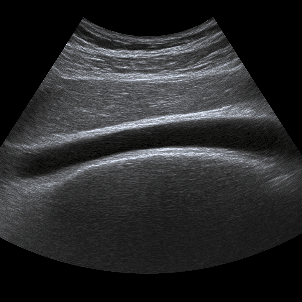
An ultrasound of the Achilles tendon is used to assess the structure, blood circulation and surrounding tissues of the Achilles tendon. The examination is performed by a specialist in radiology and provides detailed images in real time of the condition of the tendon. It can detect any inflammation, ruptures, calcifications or scarring that causes pain or impaired function in the foot.
Please note that the examination refers to one Achilles tendon. If you want to examine both Achilles tendons at the same time, you must first contact us by phone before ordering.
Ultrasound of the Achilles tendon for pain, swelling or stiffness
An ultrasound of the Achilles tendon is recommended if you experience pain, swelling or stiffness in the Achilles tendon that does not go away, especially if the symptoms affect walking, exercise or everyday movements. The examination is used to investigate both acute injuries and chronic overuse disorders.
Unlike MRI, which is mainly used to show internal structures and bones, ultrasound is a good and affordable method for examining tendons and soft tissues. The method allows the doctor to assess both structure and function in real time as the foot moves.
Common symptoms and questions
- Pain or swelling along the Achilles tendon or at the attachment to the heel bone.
- Suspected tendon inflammation (tendinitis) or overuse injury.
- Sudden pain or "snap" in the calf - suspected partial or total rupture.
- Calcifications, scar tissue or residual tenderness from a previous injury.
- Stiffness in the morning or after rest (typical of chronic tendinopathy).
- Difficulty walking on tiptoe or weakness when pushing off.
Conditions that can be detected with ultrasound of the Achilles tendon
- Tendinopathy / tendinitis – inflammation or degeneration of the tendon.
- Partial or total rupture – rupture of the Achilles tendon.
- Paratenonitis – inflammation of the membrane around the tendon.
- Calcium deposits – often with chronic overload.
- Scar formation or thickening after a previous injury.
- Bursitis – inflammation of the bursa at the tendon's attachment to the heel bone.
How an ultrasound of the Achilles tendon is performed
The examination is performed while you are lying or sitting with your foot slightly bent. A gel is applied to the skin to provide optimal contact between the probe and the skin. The doctor moves the ultrasound probe along the Achilles tendon and assesses the structure in both longitudinal and cross-sectional views. If necessary, dynamic movements can be performed to see how the tendon reacts to load.
Report and results
The images are reviewed by a specialist in radiology, who prepares a written medical report. The response is delivered digitally within a few working days and can be shared with your treating doctor or physiotherapist for further follow-up.

