


MRI Femur – Magnetic resonance imaging examination for pain, fracture or suspected skeletal disease
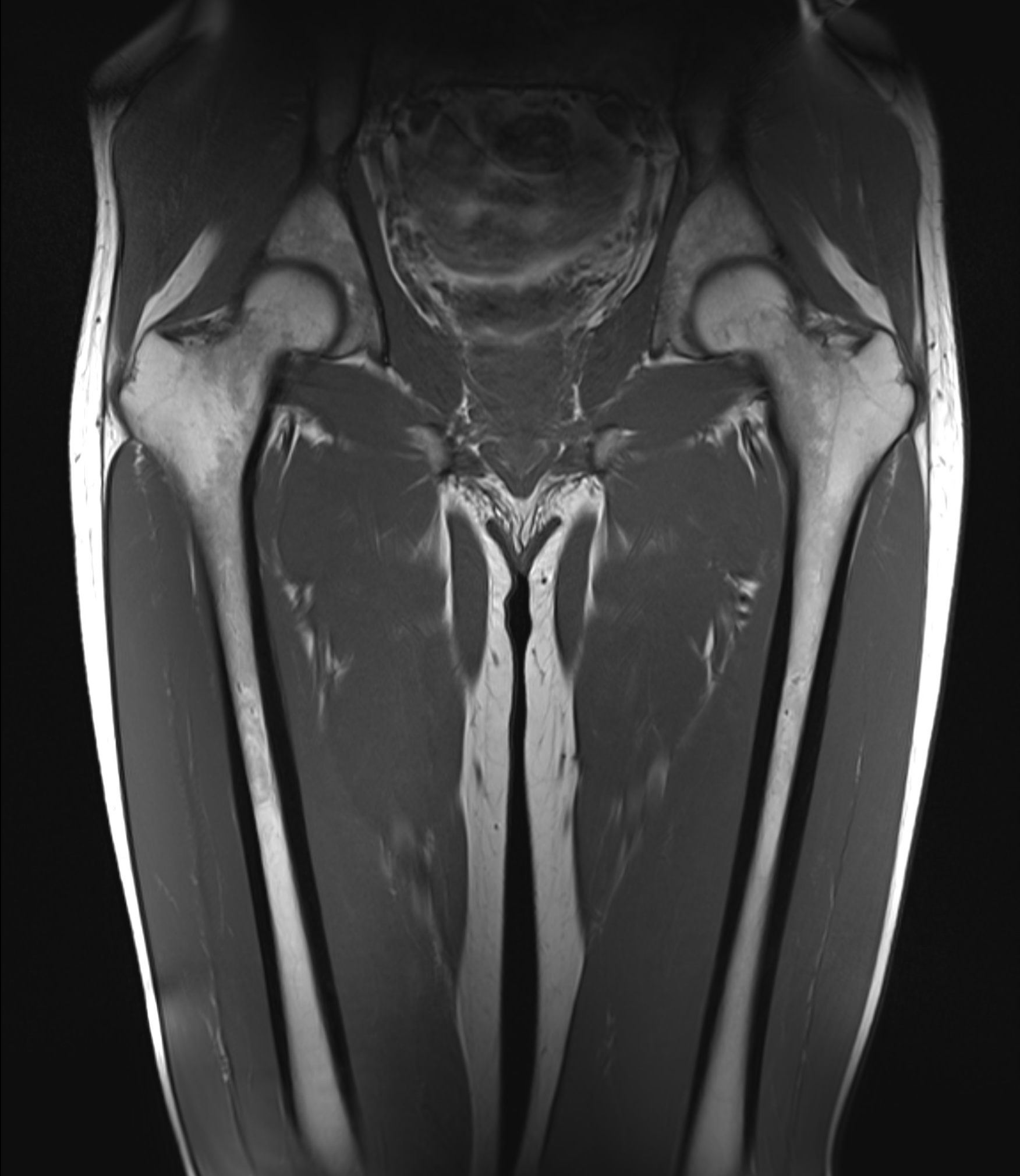
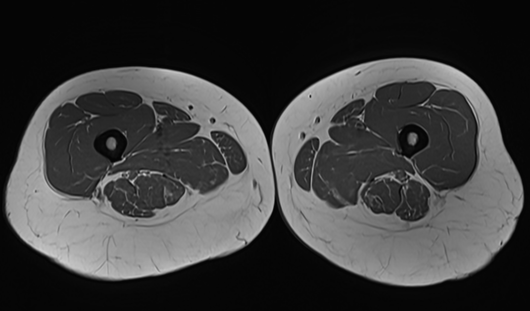
The femur (thigh bone) is the longest and strongest bone in the body – a central structure for walking, load and movement. Despite its strength, the femur can suffer from fractures, stress reactions, infections, tumors or inflammations that affect both function and quality of life. With a magnetic resonance imaging (MRI) examination of the femur, you get a very detailed picture of the skeleton, bone marrow, muscles, tendons and surrounding soft tissues. The examination is completely radiation-free, painless and particularly useful when other imaging methods have not provided sufficient information.
When is an MRI examination of the femur recommended?
MRI femur is a valuable diagnostic tool for pain that is not explained by X-ray, for suspected stress fractures, after trauma or for symptoms that indicate underlying skeletal disease. It is often used to rule out serious conditions or to map the extent of an injury.
- Unclearly painful thigh after a fall, strain or sports
- Suspected stress fracture or microfracture of the femur
- Swelling, increased heat or persistent pain in the femur
- Suspected infection in the bone or soft tissues
- Suspected tumor or metastasis in the femur
- Inflammatory disease or rheumatic changes
- Follow-up of a previous injury that has not healed as expected
MRI is particularly valuable if other examinations (e.g. plain X-ray or ultrasound) show no abnormalities, but the symptoms persist. It is also used to follow up on the healing of injuries or infections in bone tissue.
MRI is often used when the following conditions are suspected in the femur
- Stress fracture in the proximal or distal femur
- Bone marrow edema - often an early sign of overload or inflammation
- Infection in bone tissue (osteomyelitis)
- Tumors or metastases in the bone
- Inflammatory conditions - e.g. in spondylitis or vasculitis
- Tendon or muscle damage in thigh pain
- Post-traumatic changes after a previous fracture or surgery
Book an MRI of the femur - without a referral requirement
An MRI examination of the femur is an accurate way to diagnose hidden injuries or diseases in the skeleton and soft tissues. The examination is completely painless, takes approximately 20–30 minutes and does not require a referral. The images are reviewed by a specialist and a written report is delivered within a few days.
If your complaints also include the hip or knee, it may be appropriate in some cases to examine several areas at the same time. Please contact us for advice.


