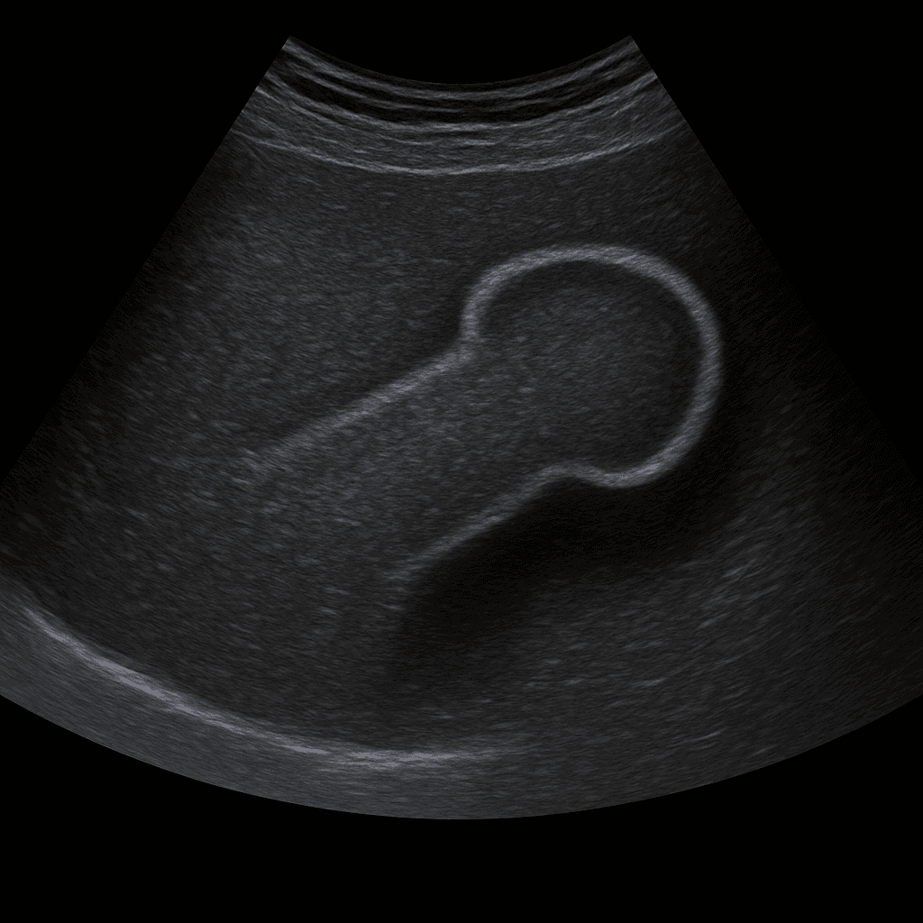
A penile ultrasound is used to examine tissue, blood flow and any structural changes in the shaft of the penis. The examination is performed by a specialist in radiology and provides real-time images that can show vascular changes, scarring, calcifications or signs of damage. Penile ultrasound is often used in cases of pain, swelling, curvature or suspected vascular involvement.
Penile ultrasound – for pain, swelling or curvature (curvature)
The examination is recommended for complaints such as pain, swelling, trauma or when the shaft of the penis has changed shape or curvature. It is also used in cases of suspicion of Peyronie's disease (plaque formation or scarring in the corpora cavernosa) and in the investigation of erection problems linked to blood flow.
Unlike MRI or CT, which are used for more extensive tissue damage or tumor investigation, ultrasound is a fast, radiation-free and effective method for assessing vascular flow and soft tissues in the penile shaft. Using Doppler technology, the doctor can analyze blood flow in the corpora cavernosa in real time.
Symptoms and questions
- Pain or swelling in the shaft of the penis.
- Bending or deformation (curvature) during erection.
- Suspected Peyronie's disease (fibrosis or plaque in the corpora cavernosa).
- Difficulty achieving or maintaining an erection (erectile dysfunction).
- Trauma, injury or suspected rupture of the corpora cavernosa.
- Hardening or calcifications under the skin.
Conditions that can be detected with ultrasound of the penis
- Peyronie's disease - scar tissue or plaque that causes bending of the penis.
- Vascular damage or impaired blood flow to the corpora cavernosa.
- Hematoma or fluid accumulation after trauma.
- Partial rupture or rupture of the corpora cavernosa.
- Inflammation or infection of the shaft of the penis.
- Calcifications or tissue changes after previous injury.
How an ultrasound of the penis is performed
The examination is usually performed while you are lying on your back. A gel is applied to the skin and the doctor moves the ultrasound probe along the shaft of the penis to assess tissue and vascular flow. The examination takes about 10–15 minutes and is completely painless. If necessary, Doppler technology is used to study blood circulation in the corpora cavernosa, sometimes after a vasodilator has been injected to simulate an erection.


