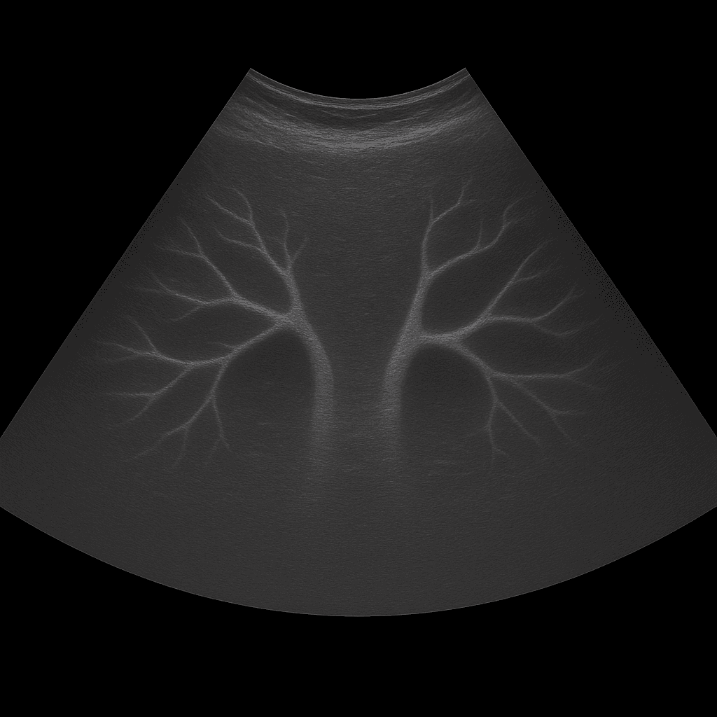
An abdominal aortic ultrasound is used to examine the body's largest artery – the aorta – in its abdominal part. The examination is performed by a specialist in radiology and provides detailed real-time images of the vessel's diameter, wall structure and blood flow. It is mainly used to detect or follow up on dilations (aneurysms), calcifications or other vascular changes that can affect blood circulation.
Abdominal aortic ultrasound – if there is suspicion of vascular dilation, a pulsating feeling in the stomach or heredity
The examination is recommended if there is suspicion of or risk of an abdominal aortic aneurysm (AAA), especially in people over 60 years of age or with a hereditary predisposition to vascular diseases. It is also used for symptoms such as a pulsating feeling in the abdomen, back pain, or as a screening in older men and women with risk factors such as smoking, high blood pressure or atherosclerosis.
Unlike MRI and CT, which are used for more advanced vascular mapping or prior to surgery, ultrasound is the first-line method for detecting and monitoring abdominal aortic aneurysms. The examination is rapid, accessible, and completely radiation-free, making it ideal for screening and follow-up over time.
Common symptoms and questions
- A throbbing sensation or bulge in the stomach.
- Pain in the stomach, back or side without a clear cause.
- A family history of aneurysm or other vascular disease.
- High blood pressure or atherosclerosis.
- Check-up after previously diagnosed abdominal aortic aneurysm (AAA).
- Follow-up after surgery or stenting in the abdominal aorta.
Conditions that can be detected with abdominal aortic ultrasound
- Abdominal aortic aneurysm (AAA) – dilation of the main artery in the abdomen.
- Calcifications in the vessel wall (atherosclerosis).
- Thrombi (blood clots) in the lumen of the vessel.
- Dissection – rupture in the vessel wall with double lumen formation.
- Narrowings or reduced blood flow in the aorta or its branches.
- Consequences after surgery or implanted vascular stent.
How an ultrasound of the abdominal aorta is performed
The examination is performed while you lie on your back. A gel is applied to the skin and the doctor moves the ultrasound probe over the upper part of the abdomen, along the iliac artery. The vessel is imaged in both longitudinal and cross-sectional views to measure its diameter and assess blood flow. In some cases, the inguinal arteries or branches to the kidneys can also be examined.
The examination is painless and usually takes 10–15 minutes. For the best image quality, you should fast for about 4 hours before the examination, as air in the intestine can impair the clarity of the image.
Order an ultrasound examination of the abdominal aorta - get a statement and recommendation from a doctor
The images are reviewed by a specialist in radiology who prepares a written medical report. The answer is delivered digitally within a few working days and can be shared with your treating doctor for further follow-up. If necessary, the findings can be followed up with MRI or CT for more detailed mapping of the vessel structure and surrounding tissues.


