MRI Small Intestine
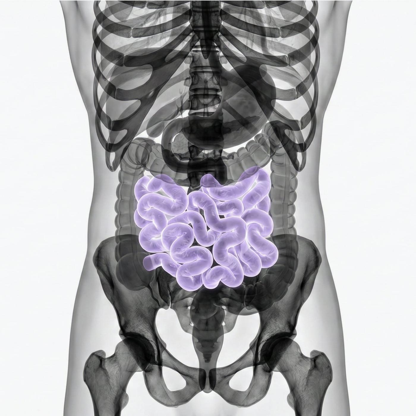
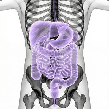
MRI small intestine is an MRI examination used to visualize the small intestine and its surrounding structures. The method is based on a magnetic resonance imaging (MRI) that uses magnetic fields and radio waves to create high-resolution images completely without radiation. The examination makes it possible to detect inflammation, strictures, tumors or other abnormalities in the small intestine and is particularly useful in chronic intestinal diseases such as Crohn's disease. In many cases, the examination is also performed with contrast media, this is included in the price but is usually decided by the radiologist based on the question.
Unlike traditional X-ray or computed tomography, MRI small intestine provides a very detailed image of both the intestinal mucosa and the surrounding tissues. The examination can show changes in the intestinal wall, bleeding, fistulas or abscesses and is often used as part of the investigation of long-term gastrointestinal symptoms. It is completely painless, radiation-free and can be combined with contrast agents for even clearer imaging.
When is MRI of the Small Intestine recommended?
MRI of the small intestine is recommended when there is suspicion of or a need to follow up on diseases of the small intestine. It is often used as a complement to colonoscopy or capsule endoscopy to obtain a complete picture of the intestinal system.

Common symptoms and complaints that may justify the examination
MRI of the small intestine is particularly valuable in cases of long-term or unexplained gastrointestinal symptoms where other investigations have not provided clear answers. The examination can help identify the underlying disease, provide evidence for correct diagnosis and guide treatment. It is often used as a complement to colonoscopy or capsule endoscopy.
- Recurrent or prolonged abdominal pain.
- Chronic diarrhea or bowel movements.
- Unexplained weight loss or malnutrition.
- Blood in the stool or suspected bleeding from the small intestine.
- Follow-up of known Crohn's disease or other inflammatory bowel disease.
- Suspected tumor or polyps in the small intestine.
Conditions when MRI of the Small Intestine may be recommended
MRI of the small intestine is used to diagnose, map and follow up various medical conditions that affect the small intestine. The examination is particularly valuable for assessing the intestinal wall and surrounding tissues, and provides important information before making decisions about medical treatment or surgery.
- Crohn's disease – inflammation of the small intestine with a risk of strictures, fistulas and abscesses.
- Tumors – detection and mapping of tumors or polyps in the small intestine.
- Bleeding – investigation of unclear anemia or suspected sources of bleeding in the small intestine.
- Stricts (stenoses) – which can cause intestinal torsion or obstruction of the passage.
- Infections or inflammation – in case of long-term stomach problems without a clear cause.
- Malformations – congenital abnormalities in the structure of the small intestine.
An MRI examination of the small intestine is a central diagnostic tool in modern gastroenterology. It makes it possible to detect both inflammatory and structural changes, and is often used as a complement when other diagnostics are not sufficient. The results are reviewed by a radiology specialist who compiles a written report for the responsible physician, which forms the basis for continued management and treatment.


