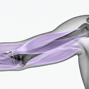
MR Brachial Plexus – examination of the nerve plexus of the arm
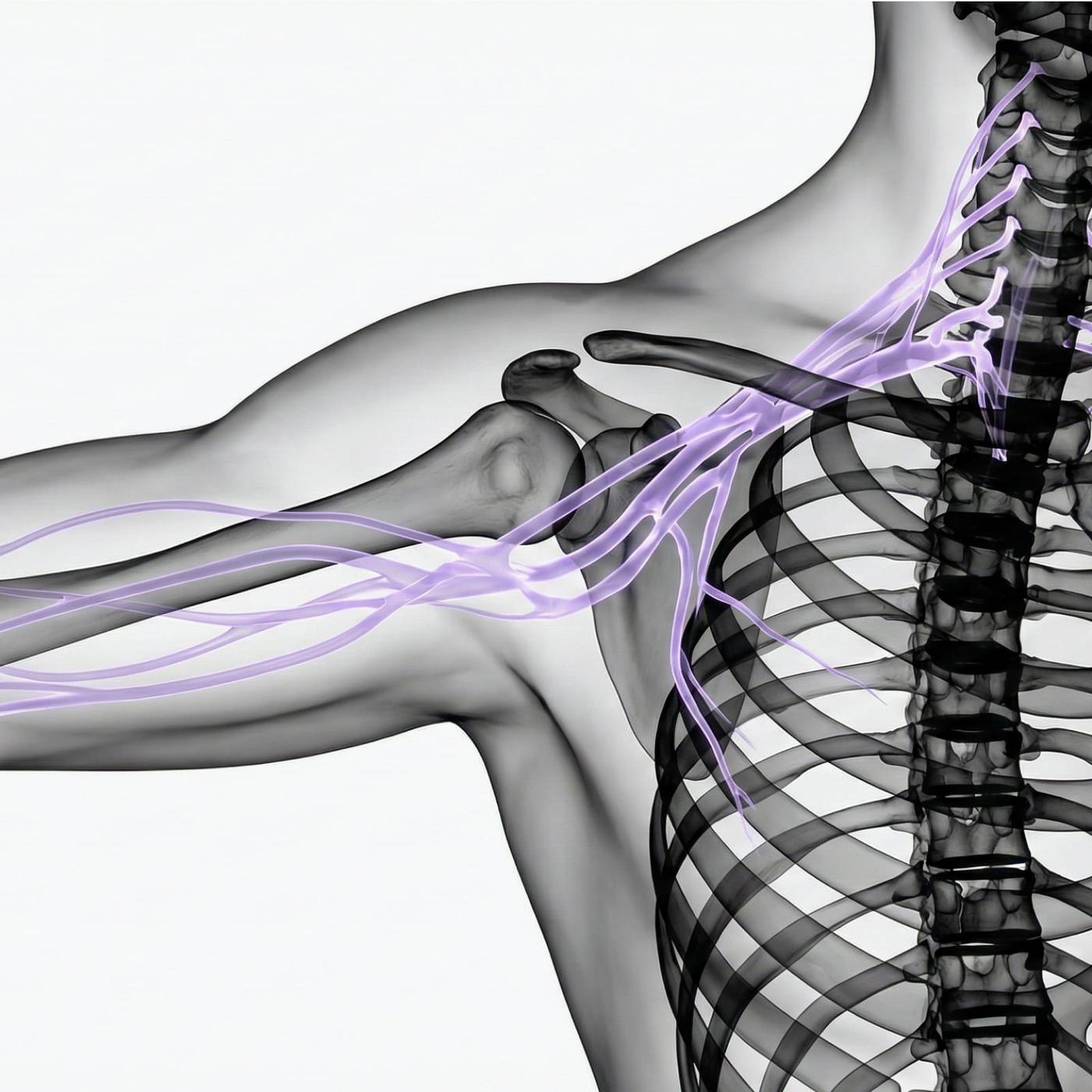
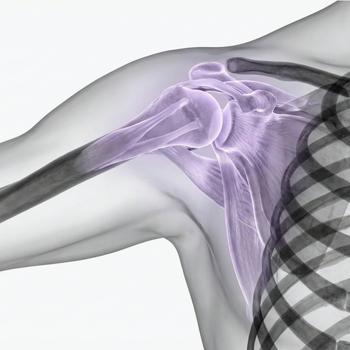
MR Brachial Plexus is an advanced magnetic resonance imaging examination that maps the large nerve plexus that originates from the spinal cord in the cervical and thoracic spine and runs down to the arm. The brachial plexus supplies the shoulder, arm and hand with both motor and sensory nerve supply. Damage or pathological changes in this area can cause pain, numbness, muscle weakness and functional impairment in the upper extremity.
The examination provides high-resolution images of nerve roots, nerve trunks and their relationship to blood vessels, muscles and surrounding tissue. The radiologist can detect interruptions in nerve pathways, nerve damage from tumors or bleeding, inflammatory processes, scar tissue or anatomical abnormalities that compress nerve structures. MRI is radiation-free, painless and a very accurate tool for investigating nerve damage in the arm.
When is an MRI of the brachial plexus recommended?
An MRI is recommended when there is suspicion of nerve damage or other effects on the brachial plexus that cannot be explained by simpler examinations. It is also used to plan treatment before surgery or to follow up on known injuries.
Common symptoms and complaints that may justify the examination:
- Severe or long-term pain in the shoulder, arm or hand without a clear cause.
- Numbness, tingling or loss of sensation in the upper extremity.
- Muscle weakness or paralysis in the arm or hand.
- Suspected nerve damage after an accident, trauma or surgery.
- Follow-up for nerve tumors or inflammatory nerve diseases.
- Investigation of symptoms in case of suspected thoracic outlet syndrome or other compression.
Conditions when MRI of the brachial plexus is recommended
- Traumatic nerve injuries – e.g. after a traffic accident, fall or sports injury.
- Postoperative changes – scar tissue or complications affecting the nerve plexus.
- Nerve tumors and metastases – which involve the brachial plexus and can cause progressive nerve damage.
- Inflammatory conditions – such as neuritis or autoimmune diseases.
- Thoracic outlet syndrome – or other conditions where nerves and vessels are pinched between ribs, muscles or vessels.
- Malformations and anatomical abnormalities – which affect the nerve supply to the arm.
- Chronic pain without a clear cause – where other investigations have not provided an answer.
Book an MRI of the brachial plexus – get a referral immediately
An MRI of the brachial plexus is a central part of the investigation of nerve damage in the arm. The examination can reveal structural damage, tumors and compression conditions that are not visible on conventional X-rays or ultrasound. By depicting nerve pathways and surrounding tissue in detail, the radiologist can provide a basis for continued medical management and targeted treatment. The results are reviewed by a specialist in radiology who compiles a report that is sent to the responsible healthcare provider. MRI is particularly valuable in cases where the symptoms are difficult to interpret or where surgical procedures are planned.


