MR Carotid Angio – mapping of the blood vessels in the neck
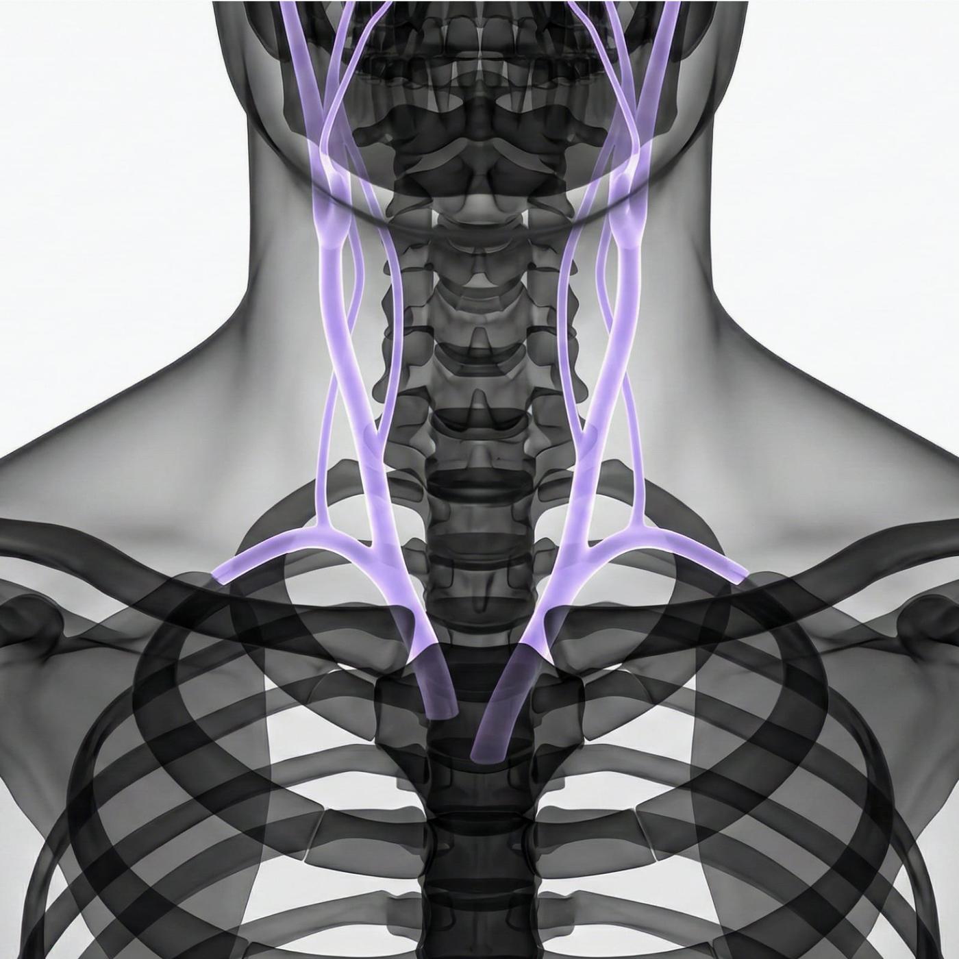
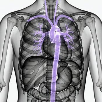
MR carotid angiography (MRA) is an advanced examination that visualizes the arteries in the neck – primarily the carotid arteries and the vertebral arteries. The method uses a powerful magnetic field and radio waves to create high-resolution images of the vascular system, completely without radiation. The examination makes it possible to detect vascular changes that can increase the risk of stroke or transient ischemic attacks (TIA).
The term angio means that the focus is on imaging blood vessels and blood flow. Unlike regular MRI, which primarily shows organs and soft tissues, MR angiography is specifically developed to produce vessel walls, narrowings and abnormalities in blood circulation.
With MR angiography, the radiologist can assess the vessel walls, blood flow and any narrowing, blockages or dilations in the carotid arteries and in the vascular segments that supply the brain. The examination is painless, radiation-free and provides very accurate images when investigating vascular disease in the central nervous system's supply.
When is MR Carotid Angio recommended?
MR angiography of the carotid arteries is recommended when there is suspicion of or follow-up on vascular diseases that affect blood flow to the brain. It is often used to investigate symptoms that may indicate impaired circulation or to follow known vascular changes before planned procedures.
Common symptoms and complaints that may justify the examination
MR carotid angiography is particularly useful for symptoms that indicate disturbances in the blood circulation to the brain. These complaints may be recurrent, long-lasting or difficult to interpret. By detecting vascular changes, the examination can help prevent stroke and other complications.
- Recurrent dizziness, fainting or balance problems.
- Sudden loss of vision or visual disturbances.
- Numbness or weakness in the face, arm or leg.
- Suspected TIA ("mini-stroke").
- Pulsating noise in the ears (tinnitus of vascular origin).
- Follow-up after previously detected vascular changes.
Conditions when MRI Carotid Angio may be recommended
The examination is used to diagnose and follow up a number of medical conditions that affect the blood vessels of the neck and blood flow to the brain. This can involve both slowly developing changes and more acute conditions. MRI angiography is particularly valuable for identifying risk factors and planning treatment.
- Carotid stenosis – narrowing of the carotid artery that can increase the risk of stroke.
- Aneurysm – bulge in a blood vessel that is at risk of rupture.
- Dissection – crack in the vessel wall that can lead to impaired blood flow.
- Thrombosis – blood clot in the arteries of the neck.
- Malformations – vascular malformations (AVM, fistulas) that affect circulation.
- Atherosclerosis – hardening of the arteries with a risk of narrowing and oxygen deficiency in the brain.
- Vasculitis – inflammation of the vessels that can damage the vessel walls and impair blood supply.
An MRI examination of the carotid arteries is a central method for detecting and mapping vascular diseases that can affect blood circulation to the brain. The examination is mainly used for planned investigations or follow-up, and can be an important complement when other imaging diagnostics have not provided sufficient information. The results are always reviewed by a specialist in radiology who compiles a written report that forms the basis for further medical treatment.

