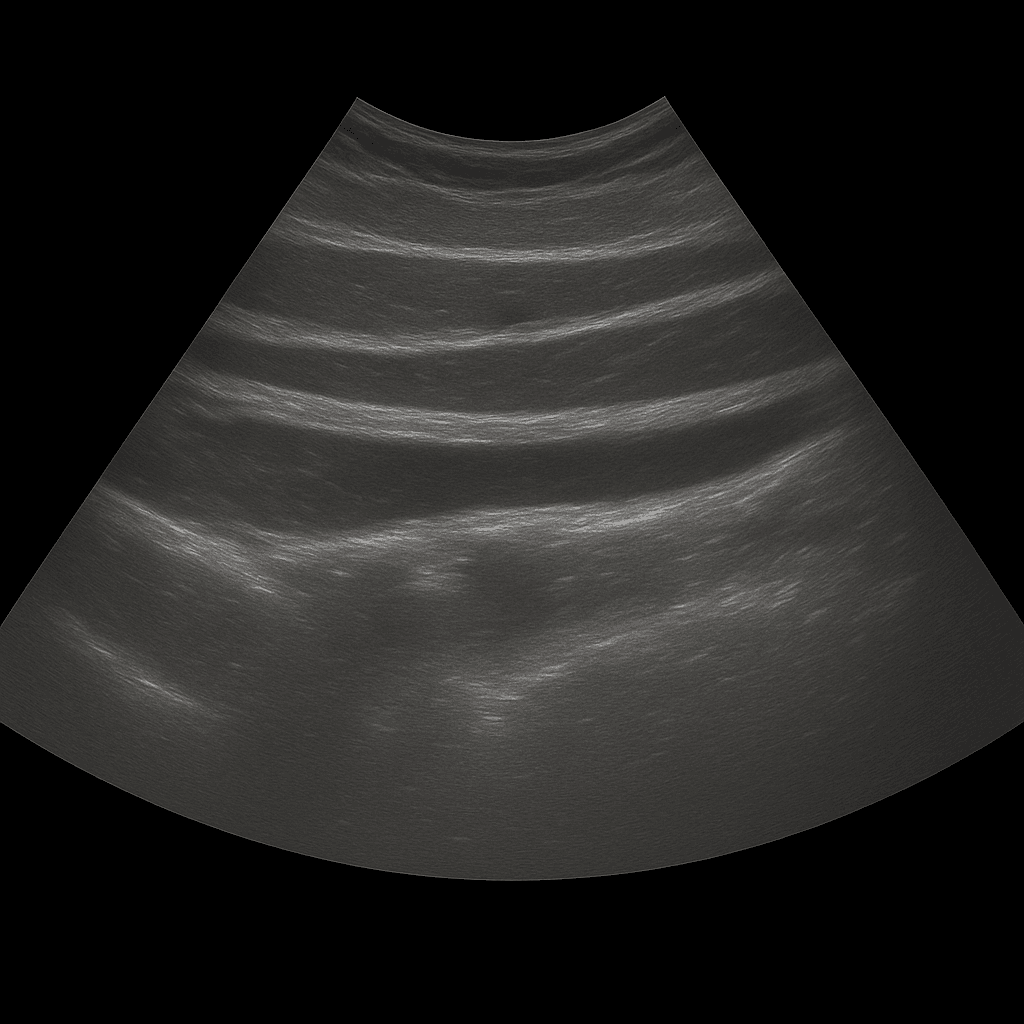
An abdominal ultrasound is used to examine the skin, subcutaneous tissue and muscles of the abdominal wall. The examination is performed by a specialist in radiology and provides detailed images in real time that can show changes that cause pain, swelling or visible bulges in the abdomen. Abdominal ultrasound is often used when there is suspicion of hernia, fluid accumulation, hematoma or other changes in the soft tissues.
Abdominal ultrasound - for pain, swelling or suspected hernia
The examination is recommended for local complaints in the abdominal wall, such as tenderness, lumps, swelling or suspicion that parts of the intestine have been pushed through the muscle wall. Ultrasound is a very reliable method for detecting and delimiting different types of abdominal wall hernias, such as umbilical hernias, scar hernias or inguinal hernias. It is also used to investigate pain after surgery, injections or trauma.
Unlike MRI and CT, which are used to map deeper organs and structures in the abdominal cavity, ultrasound is the first choice for examining superficial changes in the abdominal wall. Ultrasound shows muscles, tissue layers and possible defects in real time - without radiation or contrast media.
Common symptoms and questions
- Pain, tenderness or swelling in the abdominal wall.
- Suspected abdominal wall hernia (e.g. umbilical hernia, incisional hernia or inguinal hernia).
- Lump or bulge in the skin or subcutaneous tissue of the abdomen.
- Pain or swelling after previous surgery or trauma.
- Suspected fluid accumulation, hematoma or infection in the abdominal wall.
- Follow-up after surgery or puncture in the abdomen.
Conditions that can be detected with ultrasound of the abdominal wall
- Abdominal wall hernia – umbilical hernia, scar hernia or inguinal hernia.
- Fluid accumulation, hematoma or infection in the muscles or subcutaneous tissue.
- Scar changes and tissue defects after previous surgery.
- Fat lumps (lipomas) or other soft tissue changes in the abdominal wall.
- Inflammation or irritation of muscle and tendon attachments.
- Bulges or weaknesses in the abdominal wall after physical exertion.
How an ultrasound of the abdominal wall is performed
The examination is performed while you lie on your back. A gel is applied to the skin and the doctor moves the ultrasound probe over the area where you are experiencing discomfort. The abdominal wall is examined in different planes, both at rest and under slight tension, which makes it possible to see any hernias or defects that appear when the abdomen is loaded.
The examination is painless and usually takes 10–20 minutes. No special preparation is required. If necessary, the examination can be supplemented with MRI or CT if the change extends deeper into the abdominal cavity.
Order an ultrasound examination of the abdominal wall - get a statement and recommendation from a doctor
The images are reviewed by a specialist in radiology who draws up a written medical report. The answer is delivered digitally within a few working days and can be shared with your treating doctor for further investigation or treatment. If necessary, the findings can be followed up with other imaging diagnostics, such as MRI or CT.

