Quick version
What is an abdominal MRI?
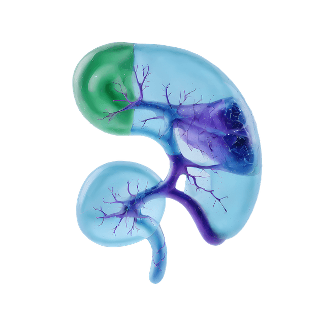
MRI (Magnetic Resonance Imaging) is a type of diagnostic imaging where strong magnetic fields and radio waves are used to create detailed images. An abdominal MRI provides high-resolution images of soft tissues and organs in the area, such as the gallbladder and bile ducts, pancreas, kidneys and urinary tract, spleen, and blood vessels. The examination is completely free from radiation and is painless.
What can an abdominal MRI show?
An abdominal MRI can reveal or confirm conditions such as:
- Tumors and suspected cancers
- Cysts (fluid-filled sacs)
- Inflammations
- Narrowing of the bile ducts or urinary tract
- Liver changes such as fatty liver or cirrhosis
- Gallstones
- Internal bleeding or fluid accumulation
- Vascular changes and blood clots
What cannot be seen on an abdominal MRI?
Certain conditions do not produce clear findings on MRI, for example:
- Functional digestive problems, such as IBS (irritable bowel syndrome)
- Temporary bowel movements and gas-related issues
- Small ulcers in the stomach or intestines (these are often examined with gastroscopy/colonoscopy)
- Allergic reactions or intolerances
How is the examination performed?
The examination is carried out at a hospital in a large tunnel-like machine with openings at both ends. You will lie still on a table that slides into the tunnel while images are taken. The examination is painless, but some people may find it uncomfortable to lie still inside the tunnel. The equipment can be noisy, so ear protectors or headphones are often used. The examination usually takes 30–60 minutes.
Treatment and follow-up
MRI is a diagnostic method and not a treatment in itself. The images are reviewed by an experienced radiologist and used as a basis for further investigation and treatment.

