Quick version
Elbow pain is not always a sign of simple overuse. When the pain radiates to the forearm, hand or fingers, nerve structures may be involved. A MRI of the elbow is then the most reliable way to establish the diagnosis and guide the treatment correctly. Through early investigation and the right intervention, long-term problems can often be avoided and normal function can be returned to more quickly.
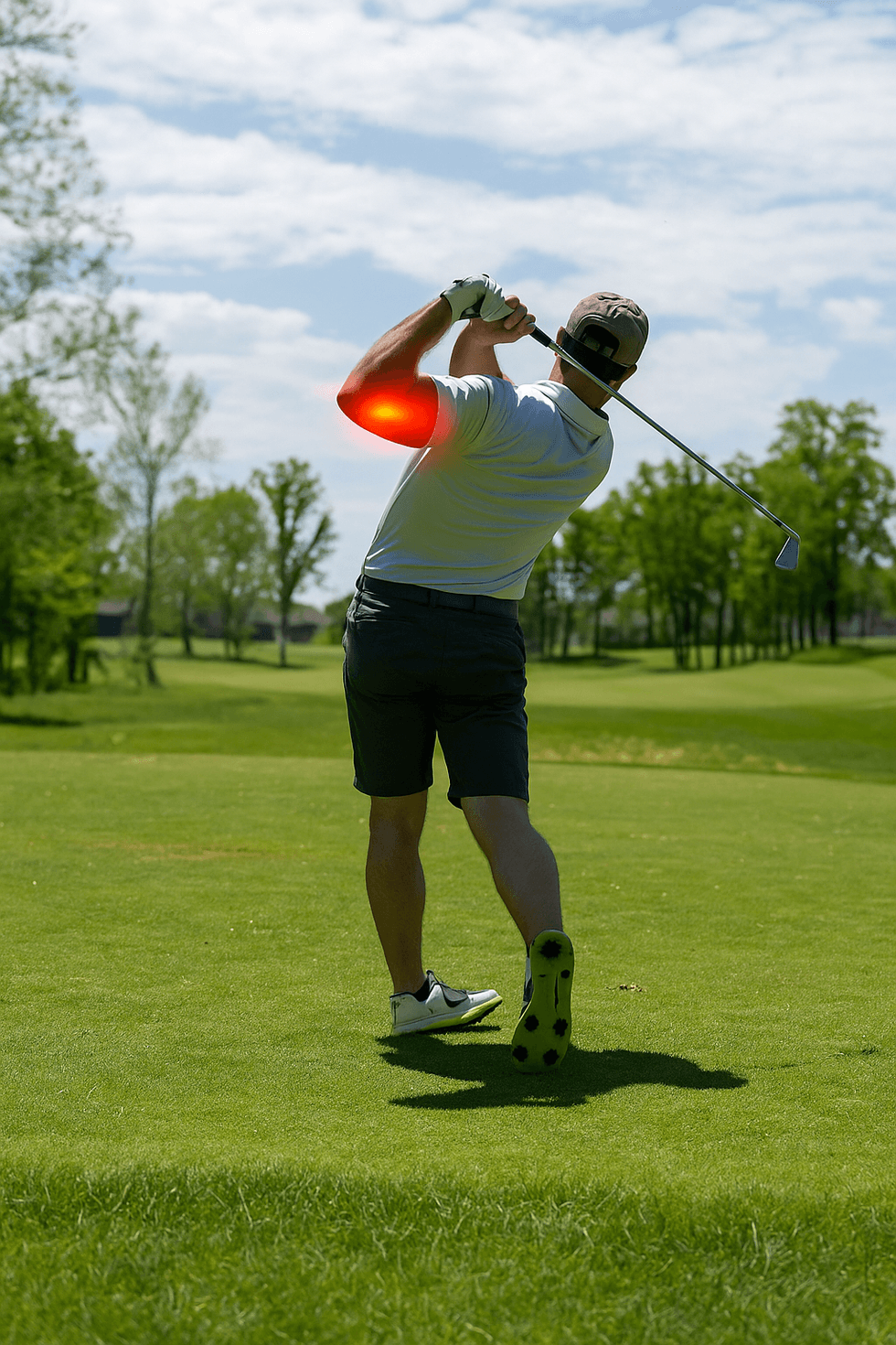
When the pain also radiates to the forearm, wrist or fingers, the cause may be a combination of tendonitis and nerve compression
Common causes of elbow pain
Elbow pain can occur for several different reasons, but one of the most common is overloading of the muscles and tendons that control the movements of the wrist and fingers. When these tissues are subjected to repeated strain, for example during strength training, golf, tennis or monotonous work at a computer, it can lead to microscopic tears in the tendon attachment. The body tries to repair the damage, but with continued strain, degenerative changes occur instead, causing the tissue to weaken and become sensitive to pain.
In golfer's elbow (medial epicondylitis), the pain is located on the inside of the elbow, where the forearm flexor muscles attach to the medial epicondyle. The condition primarily affects the flexor carpi radialis and pronator teres – two muscles that are used every time you grip, bend the wrist or rotate the forearm inwards. Typical symptoms are local tenderness, pain when gripping, weakness in the forearm and sometimes radiation along the inside of the forearm towards the wrist. The pain can be worsened by prolonged static positions, such as when working with a computer mouse or repetitive manual work.
However, not all cases of elbow pain are due to tendonitis. In many cases, the discomfort is caused or worsened by one of the major nerves in the elbow becoming pinched, which leads to tingling, numbness or burning pain. The three most important nerves that can be affected are:
- N. ulnaris: Runs behind the inside of the elbow through the cubital tunnel. When pressure or swelling occurs here, cubital tunnel syndrome occurs, which causes numbness and tingling in the ring and little fingers, pain when the elbow is bent, and reduced grip strength.
- N. radialis: Passes on the outside of the elbow and controls parts of the extensor muscles of the hand. When the nerve is pinched in the so-called radial tunnel, diffuse pain occurs over the upper part of the forearm and sometimes difficulty in extending the thumb.
- N. medianus: Runs on the front of the elbow, between the muscles of the forearm. When irritated, for example when the nerve is pinched between parts of the pronator teres muscle, it can cause tingling in the thumb, index and middle finger, as well as weakness when gripping.
The combination of overloaded tendon attachments and nerve compression means that some patients experience more extensive and long-lasting symptoms than a regular golfer's elbow would cause. Therefore, it is important to identify early whether the nerves are affected – something that often requires MRI examination of the elbow to confirm the condition.
What can an MRI of the elbow show?
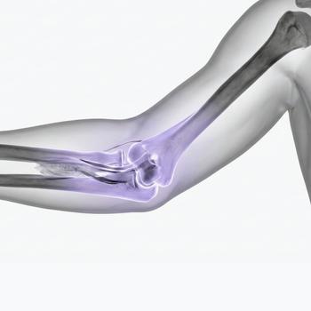
In the event of prolonged pain, suspicion of nerve involvement, or failure to improve despite rest and exercise, an MRI examination of the elbow can provide crucial information. Magnetic resonance imaging (MRI) provides detailed images of soft tissue, tendon attachments, cartilage, and nerve structures, and is used to:
- Identify tendinopathy, partial ruptures, or inflammation of tendon attachments.
- Detect swelling, fibrosis, or cysts that are pressing on nerve pathways.
- Detect signs of cubital tunnel syndrome or other nerve entrapment.
- Assess edema and changes in surrounding soft tissue following overuse.
In cases of suspected nerve compression, MRI is sometimes supplemented with neurography or electromyography (EMG). These tests measure nerve conduction and muscle activity and are used to determine where the pressure on the nerve occurs and how extensive the impact is. Neurography shows how quickly electrical signals are conducted through the nerve, while EMG records how the muscles respond to stimulation. The combination of MRI and neurophysiological tests provides a more complete picture of both the structural and functional impact in the area.
Treatment for golfer's elbow and nerve impact
In most cases, treatment begins conservatively with a focus on relieving the tissue and restoring normal function. The goal is to reduce pain, reduce any inflammation, and improve blood flow in the affected area. Treatment is tailored to the severity of the symptoms and the patient's activity level. Common elements include:
- Rest and activity modification – avoid movements that provoke pain, especially repetitive bending and gripping.
- Eccentric training – specific exercises for the forearm flexor muscles strengthen the tendon attachment and stimulate tissue healing.
- Nerve mobilization – so-called nerve gliding exercises can reduce pressure on the nerve and improve its mobility in the tissue.
- Pharmacological treatment – anti-inflammatory drugs can relieve pain and swelling. Cortisone injections can in some cases be used locally against the tendon attachment but should be given with caution.
- Rehabilitation and ergonomics – adapted training, correct lifting technique and improved working posture reduce the risk of relapse.
If the symptoms persist for more than three months despite structured rehabilitation, or if neurological symptoms such as numbness, muscle weakness or pain at night worsen, a new assessment should be made. An MRI examination of the elbow can then be supplemented with a referral to an orthopaedic or hand surgeon. If nerve compression is confirmed, surgical decompression, for example in the cubital tunnel, can reduce pressure on the nerve and give good results. The prognosis is often very good when the diagnosis is made in time and the correct treatment is initiated.
When should you seek care for long-term pain in the elbow?
Most mild elbow problems resolve with rest and self-treatment, but in some cases medical assessment is needed. You should consider further investigation if:
- The pain does not improve after rest, exercise or physiotherapy.
- You experience tingling, numbness or reduced sensation in the hand and fingers.
- You wake up at night with pain or feel reduced grip strength.
- The pain has lasted longer than two to three months despite rehabilitation.
- Previous treatment, such as cortisone injection, has not provided any improvement.
In the case of these symptoms, it is important to investigate the cause further with an MRI of the elbow to rule out nerve compression or other structural changes. Early diagnosis increases the chance of a quick recovery and reduces the risk of chronic pain.

